 |
| About Bioline | All Journals | Testimonials | Membership | News |
|
||||||
|
||||||
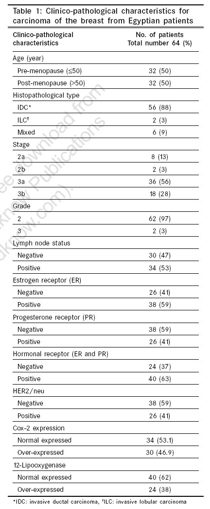
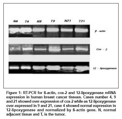
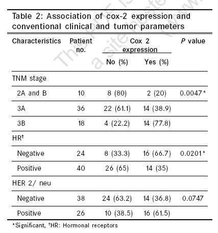
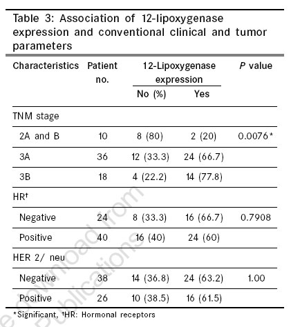
Indian Journal of Cancer, Vol. 43, No. 4, October-December, 2006, pp. 163-168 Original Article Expression of cyclooxygenase-2 and 12-lipoxygenase in human breast cancer and their relationship with HER-2/neu and hormonal receptors: Impact on prognosis and therapy Mohammad AM, Abdel HA, Abdel W, Ahmed AM, Wael T, Eiman G Biochemistry and Molecular Biology Unit, Departments of Cancer Biology, NCI, Cairo University Code Number: cn06026 Abstract Background: A number of studies have shown over-expression of cox-2 in breast cancer. Also it has been recorded that human breast cancer expresses high level of cox-2 and 12-lipoxygenase which may be beneficial in future therapy plan for those patients.Aims: The present study aims to examine the level of transcripts of cox-2 and 12-lipoxygenase in Egyptian breast cancer patients and to compare between the expressions of both enzymes and TNM staging, hormone receptors status (including estrogen and progesterone) and HER2/neu expression. Materials and Methods: Total cellular RNA was extracted from 64 frozen tissue samples of breast carcinoma and their corresponding normal adjacent tissues. Cox-2 and 12-lipooxygenase expressions were detected using RT-PCR. Hormonal receptors as well as HER2/neu were detected immuno-histochemically for each patient. Results: About 47 and 62.5% of carcinoma samples showed over-expression of cox-2 and 12-lipooxygenase respectively as compared to their corresponding normal tissues. The results revealed that cox-2 significantly associated with TNM staging ( P =0.0047) and hormonal receptors status ( P = 0.0201). The relationship between cox-2 and HER2/neu expression was close to a significant value ( P =0.0747). 12-lipooxygenase showed only significant association with TNM staging ( P =0.0076). Neither hormonal receptors nor HER2/neu showed significant association with this enzyme. Conclusion: Elevated levels of cox-2 and 12-lipoxygenase expression were detected in human breast cancer. Also, the results revealed that cox-2 and 12-lipooxygenase mRNA expressions are associated with TNM staging in human breast cancer. Furthermore, there is an inverse association between cox-2 expression and hormonal receptor status. This observation may drive us to the possible role of those two enzymes in determining the plan of therapy of breast cancer patients. Keywords: Breast cancer, cox-2; estrogen receptor, Her-2/neu, lipoxygenase-12, progesterone receptor Cyclooxygenase is a rate-limiting enzyme for prostaglandin (PG) synthesis from arachidonic acid. It incorporates two molecules of oxygen into arachidonic acid and produces PGG2 which is reduced by the same enzyme to PGH2.[1],[2],[3] Two isoforms of the same enzyme are known; cyclooxygenase-1, which is a constitute enzyme expressed in many tissue and cyclooxygenase-2, which is an inducible enzyme synthesized at inflammatory lesions by cytokines, growth factors and tumor promoters.[4],[5] In breast cancer, greater amounts of prostaglandins are produced in tumor tissues than normal tissue derived from the same organ.[6] Increased level of cyclooxygenase-2 in human breast cancer cell has been reported, denoting that breast cancer tumor cells have more cyclooxygenase-2 protein than normal adjacent cells.[7] Cyclooxygenase-2 is also involved in angiogenesis process,[8],[9] by way of regulating the vascular epithelial growth factor pathway and Peroisome proliferators-activated receptor (PPARg).[8],[10] However, the involvement of cox-1 and cox-2 in the progression and metastasis of breast cancer is not yet fully understood, although it was mentioned that over-expression of cox-2 in breast cancer cells increased metastatic potential.[11],[12],[13] The current study examined the level of the transcripts of cox-2 and 12-lipoxygenase in human breast cancer and examined the relationship between these two enzyme and clinical outcomes in those patients which may be important in the plan of therapy. Materials and Methods Patients This study was performed on tissue samples from 64 women aged between 27-74 years (median 47.6 years) who were examined during 2000-2003 at the hospital of National Cancer Institute, Cairo University. Breast cancer tissues and their corresponding matched adjacent normal tissues were obtained immediately after surgery. Tissues were frozen in liquid nitrogen within one hour of surgical removal and subsequently stored at -80°C. All tissues analyzed were obtained from patients who had provided informed consent and with Institutional Review Board approval in accordance with the NCI, Cairo University. All clinicopathological characteristics for the cases under investigation were collected from epidemiology and biostatistics department and are summarized as shown in [Table - 1]. Specimen preparation Formalin-fixed, paraffin-embedded blocks were obtained from the primary breast tumors received for frozen section examined at the Department of Pathology of the National Cancer Institute, Cairo. A slide stained with hematoxylin and eosin was prepared from each block and used for pathological confirmation of breast cancer. For the same block, 4-micron tissue sections were prepared for immunohistochemical study. The two pathologists among the authors reviewed the slides according to the world health organization (WHO) classification of breast tumors. The histological grading system was employed according to the Bloom, Scarff and Richardson grading system. Immunohistochemical analysis Immunohistochemical reactions were performed using the following mouse monoclonal antibodies applied for one hour at room temperature (all from DakoCytomation, Denmark): Hercep Test kit for c-erbB-2 (HER2/neu), clone 1D5 for estrogen receptor (ER) and 1A6 for progesterone receptor (PgR) (optimally prediluted) The antigens were visualized using biotinylated antibodies, streptavidin-peroxidase complex (LSAB2, DakoCytomation) and 3,3′-diaminobenzedine (DAB) (DakoCytomation). Detection of HER2/neu, ER and PgR expression was preceded by 15-min exposure of the sections to boiling in antigen retrieval solution (Dako Cytomation) in a microwave oven. Appropriate positive controls were used. For negative controls, primary antibodies were omitted. Cases were considered positive for ER and PgR when they showed a nuclear staining in more than 5% of tumor cells. Evaluation of c-erbB-2 over-expression was done according to the manufacturer′s instructions by scoring the percentage of stained invasive malignant cells and tissue was considered to be positive for c-erbB-2 expression if it had a score of 2+ or 3+ and as negative if it had a score of 0 or 1+. RT-PCR Total RNA from the tissues collected was extracted using high pure RNA tissue kit (Roche, Diagnostics GmbH, Germany) according to the manufacturer′s instruction and RNA was dissolved in diethylpyrocarbonate (DEPC) water and stored at -70°C until assaying. Routine reverse transcriptase polymerase chain reaction (RT-PCR) was carried out using a PCR mix that was commercially available (Qiagen One-step RT-PCR kit, Germany). The primers used were: for cycloxygenase-2: 5′ tgtatgagttgtggatttga 3′ and 5′ tggatggggtttcagaaata 3′, for 12-lipoxygenase: 5′ tcaactggagaaaagaacttca 3′ and 5′ gatcacgttggctggaat 3′ and for β-actin: 5- acactgtgcccatctacgagg 3′ and 5′ aggggccggactcgtcatact 3′. Reactions were carried out at the following conditions: a reverse transcription reaction temperature of 50°C for 30 min. followed by initial PCR activation step at 95°C for 15 min. followed by 35 cycles consisted of 94°C for one min, 56°C for one min and 72°C for one min and final extension at 72°C for 10 min. PCR products were separated on a 2% agarose gel and the intensity of bands were measured using BioDocAnalyzer (BioMetra, Germany). Statistical analysis Statistical analysis was performed using statistical package for social sciences (SPSS) software for PC (version 10). Chi-square test was used to determine whether there was a statistically significant difference in the expression of cox-2 and 12-lipoxygenase between the cancer specimen and the corresponding adjacent normal specimen. Also, Chi-square test was used to analyze the statistical correlation between the over-expression of cox-2 and 12-lipoxygenase with various clinicopathological factors. P -value less than 0.05 was considered statistically significant. Results In this study cox-2 expression as well as 12-lipoxygenase were detected using reverse transcriptase polymerase chain reaction (RT-PCR) in 64 human breast carcinomas and their corresponding normal adjacent tissues. Bands obtained were analyzed using BioDocAnalyzer (Biometra, Germany). Cox-2 or 12-lipoxygenase were defined positive when their percent of intensity was greater by 5% than the control.[18] Hormonal receptors (including ER and PgR) and HER2/neu were detected immunohistochemically for each patient. By RT-PCR analysis, 30 of the 64 (46.9%) cancer specimens showed over-expression of cox-2 as compared to their corresponding normal adjacent tissues, [Figure - 1] shows representative samples for cox-2 over-expression of breast cancer tissues as compared to their normal tissues. The association of cox-2 and TNM stage was statistically analyzed. There was a steady significant increase of cox-2 expression with increased TNM staging scoring 20% in both 2A and 2B whereas it increased to 38.9% and 77.8% in 3A and 3B respectively ( P =0.0047). The immunohistochemical analysis reveled that 63% of the patients exhibited positive hormonal receptors including ER and PgR. The correlation between cox-2 expression and the hormone receptor status revealed that 33.3% of the negative hormone receptor patients exhibited normal cox-2 expression, while 66.7% of the negative hormone receptor cases exhibited cox-2 over-expression. It also found that 65% of the positive hormone receptor patients has normal cox-2 expression while the rest (35%) showed cox-2 over-expression. This association was statistically significant ( P = 0.0201). Forty one percent of the cases examined exhibited over-expression of HER2/neu, 61.5% of them showed over-expression of cox-2 while in HER2/neu negative cases (about 59% of all breast cases studied), cox-2 over-expression was detected in 36.8% of them. However, the statistical analysis is close to a significant relationship ( P =0.0747) between cox-2 expression and HER-2/neu patient′s status. [Table - 2] summarizes these associations between cox-2 expression with TNM staging, HR and HER2/neu. By RT-PCR analysis, 24 of the 64 (38%) cancer specimens showed over-expression of 12-lipoxygenase as compared to their corresponding normal adjacent tissues, [Figure - 1] shows over-expressions in cases 9 and 21 while it was normal in case 4 as compared to their normal tissues. The association of lipoxygenase-12 and TNM stage were statistically analyzed. There was a steady significant increase of lipoxygenase-12 expression as in cox-2 with increased TNM staging where it scored 20% in both 2A and 2B and increased to 66.7% and 77.8% in 3A and 3B respectively. This association was statistically significant ( P =0.0076). The relation between lipoxygenase-12 expression and the hormone receptors status revealed that 33.3% of the negative hormone receptor patients exhibited normal lipoxygenase-12 expression, while 66.7% of the negative HR cases exhibited lipoxygenase-12 over-expression. It was also found that 40% of the positive HR patients has normal lipoxygenase-12 expression while the rest (60%) showed lipoxygenase-12 over-expression. This association was statistically non-significant ( P =0.7908). Forty one percent of the cases examined exhibited over-expression of HER2/neu, 61.5% of them showed over-expression of lipoxygenase-12 while in HER2/neu negative cases (about 37.5% of all breast cases studied), lipoxygenase-12 over-expression was detected in 63.2% of them. However, the statistical analysis revealed no significant relationship ( P =1.0) between lipoxygenase-12 expression and HER-2/neu patient′s status. [Table - 3] summarizes these associations. The relationship between both enzymes studied was also analyzed. From 24 patients that normally expressed 12-lipoxygenase, only 10 of them showed over-expression of cox-2, while 20 patients out of 40 that showed over-expression of 12-lipoxygenase expressed high level of cox-2. The statistical analysis showed no significant correlation between both enzymes ( P = 0.726). Discussion Elevated cox-2 expression in breast carcinoma was associated with large tumor size, high histological grade, negative hormone receptors status, high proliferation rate, high p53 expression and the presence of HER-2 oncogene amplification.[19] Jiang and his colleagues[20] showed that human breast cancer aberrantly expresses high levels of cox-2 and 12-lipoxygenase that may have prognostic value in patients with breast cancer. Our study on 64 samples from breast carcinoma patients confirms previous reports. In our series, cox-2 was positively expressed in 47% of the 64 studied cases with negative detectable cox-2 expression in normal mammary gland epithelium surrounding malignant tissue. This observation is in keeping with recent data showing frequently higher cox-2 expression in various epithelial neoplasia compared with adjacent normal tissue.[21],[22],[23],[24] Also the expression of cox-2 was correlated with other tumor variables such as TNM staging, HR and HER-2/neu status. It was observed that there was a steady significant increase of cox-2 expression with increased TNM staging. A significant inverse association between over-expression of cox-2 and hormone receptor was detected, however the study found a close to significant correlation between the expression of cox-2 and HER-2/neu, in spite of an increase of the cox-2 expression among the patients with positive Her-2/neu expression. Also an increase of the cox-2 expression in the patients showing positive HER2/neu was noticed. These findings are supported by results reported by Ristimaki et al[22] and Sucic et al[25] indicating that elevated cox-2 expression is more common in breast cancer with poor prognostic characteristics and is associated with an unfavorable clinical outcome. Subbaramaiah and colleagues[26] showed that a high level of cox-2 protein was detected in 14 of 15 breast cancer samples that were positive for Her2/neu expression. In contrast, the level and frequency of cox-2 expression were significantly lower in HER2/neu negative samples, as only four of 14 of these samples were positive for cox-2. The mechanism by which cox-2 is up-regulated in breast cancer is unknown, but one possibility is that cancer cells become intrinsically more active in expressing cox-2 than do the non-neoplastic cells. To this end, both inactivation of tumor suppressor genes, such as p53 and activation of oncogenes, such as HER2/neu, have been implicated in induction of cox-2 expression.[27] Our results support this hypothesis, because elevated cox-2 expression was more common in samples with amplification of HER2/neu. However, because elevated cox-2 expression was not restricted to HER2/neu positive tumors, several other factors (such as activated Ras, over-expressed Src or epidermal growth factor receptor pathway) are likely to be responsible for elevated cox-2 expression as well.[27] Howe et al[28] derived a genetic evidence that cox-2 contributes to HER2/neu induced mammary tumorigenesis. Several studies have suggested that fatty acids and other enzymatically oxidized lipids may play significant role in the development and progression of breast cancer. However, few studies have examined the molecular regulation of the enzymes involved in the biosynthesis of fatty acid derived metabolites in breast cancer cells and tissues.[29] 12-HETE (Eicosanoid product derived from archidonic acid through 12-lipoxygenase pathway) has been shown to increase growth, proliferation and metastatic potential.[30] In the present study, we have shown that 12-lipoxygenase expression was up-regulated to some extent (38%) in breast cancer tissues, compared with their normal counterparts. These results are in agreement with Jiang et al[20] who reported increased levels of 12-lipoxygenase in breast cancer as compared with control tissues. The increase of 12-lipoxyenase expression in breast cancer tissues would be expected to result in increase of 12-HETE production and to increase metastatic potential which would be reflected in the clinical outcome of patients (not significantly apparent in this study). Our data revealed significant correlation between levels of 12-lipooxygenase and TNM staging, however it was insignificant with both hormone receptors and HER-2/neu. We have also analyzed the relationship between cox-2 and 12-lipoxygenase expression. Unfortunately, no significant correlation existed between the two enzymes. Conclusion Our findings are in accordance with the results of others in that they indicated elevated cox-2 and 12-lipooxygenase expression in breast carcinoma. Also, the results revealed that cox-2 and 12-lipooxygenase mRNA expressions are associated with TNM staging in human breast cancer. Furthermore, an inverse association between cox-2 expression and hormonal receptor status was detected. This observation may drive us to the possible role of those two enzymes in determining the plan of therapy for breast cancer patients.References
Copyright 2006 - Indian Journal of Cancer The following images related to this document are available:Photo images[cn06026t2.jpg] [cn06026t1.jpg] [cn06026f1.jpg] [cn06026t3.jpg] |
| |||||||||
