
|
Indian Journal of Cancer
Medknow Publications on behalf of Indian Cancer Society
ISSN: 0019-509X EISSN: 1998-4774
Vol. 46, Num. 3, 2009, pp. 194-202
|
Indian Journal of Cancer, Vol. 46, No. 3, July-September, 2009, pp. 194-202
Original Article
Clinical significance of matrix metalloproteinase 2 and 9 in breast cancer
Shah FD, Shukla SN, Shah PM, Shukla HK, Patel PS
Biochemistry Research Division, The Gujarat Cancer and Research Institute, Ahmedabad - 380 016
Correspondence Address:Biochemistry Research Division, The Gujarat Cancer and Research Institute, Ahmedabad - 380 016
prabhudas_p@hotmail.com
Code Number: cn09043
PMID: 19574670
DOI: 10.4103/0019-509X.52953
Abstract Background: Invasion and metastasis are the most strenuous problems in the management of breast cancer. These events require diverse proteolytic enzymes, among which MMP-2 and MMP-9 play a significant role in degradation of type IV collagen, the major component of the basement membrane. Therefore, the major objective of the study is to evaluate the clinical usefulness of MMP-2 and MMP-9 with respect to malignant tumor growth, invasion, and metastasis in breast cancer.
Materials and Methods: Gelatin zymography was performed on 157 tissue extracts of malignant and adjacent normal breast tissues as well as negative and positive lymph nodes from 49 breast cancer patients. Statistical analysis was carried out using SPSS statistical software (version 10).
Results: ProMMP-2 levels were significantly higher in adjacent normal tissues. Active MMP-2 and MMP-9 levels were higher in malignant breast tissues. Activation ratios of MMP-2 and MMP-9 were significantly higher in malignant breast tissues and in patients with lymph node metastasis. ProMMP-2, active MMP-2, and active MMP-9 could significantly discriminate between malignant and adjacent normal breast tissues. The MMP-2 activation ratio showed significant discriminatory efficacy between patients with and without lymph node metastasis and significant association with increased risk of lymph node metastasis in node-negative patients.
Conclusion: The results indicate significant clinical utility of these proteolytic enzymes in malignant tumor growth, invasion, and metastasis in breast cancer.
Keywords: Breast cancer, MMP-2, MMP-9, zymography
Introduction
Breast cancer, a pathologically and clinically heterogeneous disease is the most frequent malignancy among women. [1] It is a malignancy with variable prognosis as local invasion into the surrounding tissues and subsequent spread to distant organs are very common features of malignant breast tumors. The metastatic process involves intravasation and extravasation of tumor cells, followed by reimplantation of tumor cells, formation of a new tumor stroma, and neoangiogenesis, to consolidate a secondary tumor at a distant site. [2] These steps in the promotion of malignant tumors require the involvement of proteolytic enzymes, which can degrade the protein components of the extracellular matrix (ECM).
The degradation of ECM and components of the basement membrane by proteases, facilitates the detachment of tumor cells, their crossing of tissue boundaries, and invasion into adjacent tissue compartments. There are several classes of proteolytic enzymes involved in ECM degradation. Recently, the importance of tumor-associated proteases in invasion and metastasis has been demonstrated in a variety of solid malignant tumors. Matrix metalloproteinases (MMPs), a class of proteolytic enzymes, are thought to play a central role in this process, in view of their ability to degrade many ECM components and their substances. [3] At least 22 members of the human MMP gene family have been identified. Among these, gelatinases comprised of gelatinase A (MMP-2) and gelatinase B (MMP-9), are associated with the malignant phenotype of tumor cells, because of their unique ability to degrade the type-IV collagen, a major component of the basement membrane. The gelatinases are secreted as zymogens and cleave to the active form and their function is tightly regulated by several different mechanisms. [3],[4] Numerous investigators have reported the clinical significance of MMP-2 and MMP-9 in breast cancer. [5],[6],[7] Therefore, the purpose of the present study is to measure the zymogen, as well as the active forms of MMP-2 and MMP-9 in extracts of malignant and adjacent normal breast tissues and in positive and negative lymph node tissues. These parameters have been investigated to assess their clinical usefulness with respect to malignant tumor growth, invasion, and metastasis, in breast cancer patients.
Materials and Methods
Tissue samples
Forty-nine breast cancer patients who underwent surgery as the primary treatment were included in the study [Table - 1]. Among these, a majority of the patients (79.6%) were with invasive ductal carcinoma, histological type. Patients with other (20.4%) histopathology types includes three patients with infiltrating ductal carcinoma, three patients with medullary carcinoma, two patients with ductal carcinoma in situ, and one patient each with papillary carcinoma and tubular carcinoma. Staging was done according to TNM (Tumor, Node, Metastasis) staging and the patients were grouped into early stage (stage I and II) and advanced stage (Stage III and IV) of the disease. According to the lymph node involvement, the patients were grouped into patients with lymph node involvement (positive) and without lymph node involvement (negative). The study was approved by the institutional ethics committee. A total of 157 tissue samples were collected from these patients at the time of surgery. It included 48 malignant tissues, 49 adjacent normal tissues, 36 lymph node tissues positive for malignancy, and 23 lymph node tissues negative for malignancy. Malignant and adjacent normal breast tissues and positive and negative lymph node tissues were homogenized (1 gm /10 ml) on ice with 0.1 M phosphate buffer saline (pH = 7.4) using glass mortar and pastel. The lysates were centrifuged at 15,000 rpm in a cooling centrifuge for 30 minutes. The supernatant was used for gelatin zymography. Protein contents were estimated from the supernatant by the Lowry method. [8] Tissue lysates equivalent to 50 µg protein were mixed with equal volumes of sample buffer (4% SDS, 20% Glycerol, 0.004% Bromophenol, 0.125 M Tris HCL) and kept at room temperature for 30 minutes. Gelatin zymography was performed according to the method of Lorenzo et al., [9] with the necessary modifications. Briefly, the samples were run under nondenaturing and nonreducing conditions at a constant voltage (100 V) until the dye front reached the end of the plate. After electrophoresis, the gels were washed twice with 0.25% Triton-X-100 (100 ml) for 15 minutes each. The gels were then incubated overnight in an activation buffer. Next day, the activation buffer was decanted and the gels were stained with 0.1% coomassie brilliant blue R-250, in 40% propanol, for a minimum of 1 hour. The gels were then destained in 7% acetic acid. Proteolytic activity was represented by clear lysis bands of degraded protein on a uniformly blue background. Gels were quantitated using the Bio-Rad gel documentation system. The relative proteinase activity was determined for each proteinase by multiplying the area of each lysed band by its optical density (ODFNx01mm 2 ).
Statistical analysis
Data were statistically analyzed using the SPSS statistical software (version 10). Student′s unpaired ′t′ test and paired ′t′ test were used to compare the mean levels of MMP-2 and MMP-9 in malignant and adjacent normal breast tissues and positive and negative lymph node tissues. Receiver′s Operating Characteristic (ROC) curves were constructed to determine the discriminating efficacy of MMP-2 and MMP-9 between malignant and adjacent normal breast tissues. ROC curves were plotted to find out the discriminatory efficacy of the activation ratio of MMP-2 and MMP-9 between patients with and without lymph node metastasis. Multivariate analysis was carried out to correlate the markers with clinicopathological parameters including age, menopausal status, lymph node involvement, early and advanced stage of the disease, BR score and nuclear grade of the tumor. Odds ratio (OR) was calculated to assess the association of the activation ratio of MMP-2 and MMP-9 with the risk of developing lymph node metastasis, in lymph node negative patients. Values of the parameters were expressed as Mean ± S.E. "p" values less than 0.05 were considered statistically significant.
Results
Expression of matrix metallo proteinases by substrate zymography
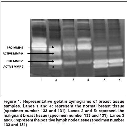
Gelatin zymography identifies and separates the gelatinases, MMP-2, and MMP-9 in both latent and active forms. [Figure - 1] shows the representative patterns of the gelatin zymogram of various tissue samples. In the Figure, lanes 1 and 4 represent MMP-2 and MMP-9 expression patterns in adjacent normal breast tissues, lanes 2 and 5 represent malignant breast tissues, and lanes 3 and 6 represent positive lymph node tissues. Zymograms showed distinct separation of the active forms and latent forms of MMP-2 and MMP-9 in the tissue samples.
Pro, active, total, and activation ratio of MMP-2 and MMP-9 in malignant and adjacent normal breast tissues
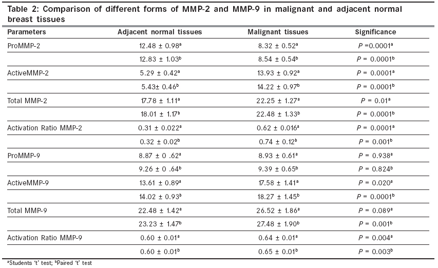
[Table - 2] shows the comparison of the mean levels of pro MMP-2, active MMP-2, total MMP-2 (pro MMP-2 and active MMP-2), activation ratio of MMP-2 (active MMP-2 / total MMP-2), pro MMP-9, active MMP-9, total MMP-9 (ProMMP-9 and activeMMP-9) and activation ratio of MMP-9 (active MMP-9 / Total MMP-9) in malignant and adjacent normal breast tissues obtained by student′s unpaired ′t′ test and paired ′t′ test. In the malignant tissues, the mean levels of proMMP-2 were significantly lower ( P =0.0001) as compared to the adjacent normal tissues. The mean levels of active MMP-2, total MMP-2, and activation ratio of MMP-2 in the malignant tissues were significantly higher as compared to the adjacent normal tissues ( P =0.0001 , P =0.01 , and P =0.0001; respectively). The mean levels of active MMP-9, total MMP-9, and activation ratio of MMP-9 were higher in malignant tissues as compared to adjacent normal tissues. Further, the differences in active MMP-9 and activation ratio of MMP-9 levels were also statistically significant ( P =0.020, P =0.004; respectively). Pro MMP-9 levels were comparable between malignant and adjacent normal breast tissues. Paired ′t′ test also showed that pro MMP-2 levels were significantly lower in malignant tissues as compared to adjacent normal breast tissues ( P =0.0001). The mean levels of active MMP-2, total MMP-2 and activation ratio of MMP-2 in the malignant tissues were significantly higher as compared to the adjacent normal tissues ( P =0.0001, P =0.0001 and P =0.001; respectively). Pro MMP-9 levels were comparable between malignant tissues and adjacent normal breast tissues. The mean levels of active MMP-9, total MMP-9 and activation ratio of MMP-9 in the malignant tissues were significantly higher as compared to the adjacent normal tissues ( P =0.0001, P =0.001 and P =0.003; respectively).
Activation ratio of MMP-2 and MMP-9 in patients′ lymph node tissues
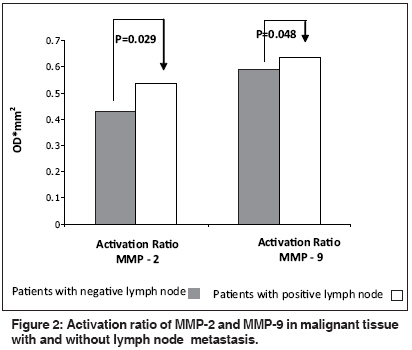
[Figure - 2] shows activation ratio of MMP-2 and MMP-9 in patients with and without lymph node metastasis. Activation ratio of MMP-2 and MMP-9 was significantly elevated in malignant tissues of patients with lymph node metastasis as compared to malignant tissues of patients without lymph node metastasis ( P =0.029 and P =0.048, respectively).
ROC curve analysis
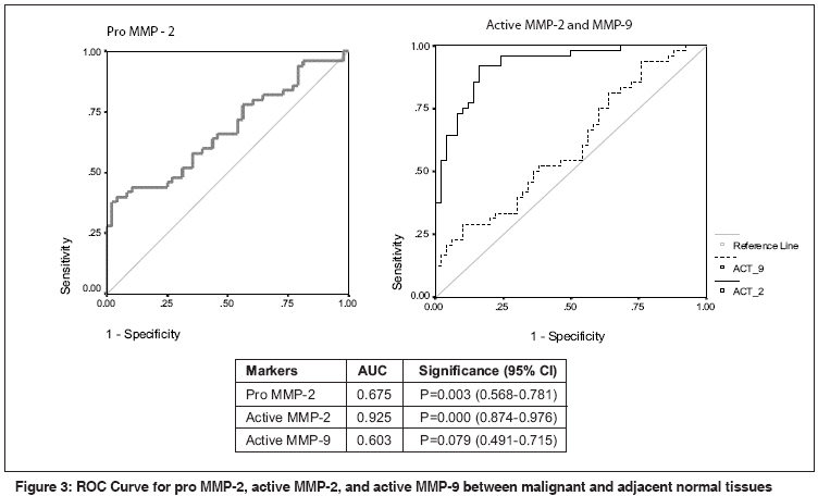
[Figure - 3] shows ROC curves for pro MMP-2, active MMP-2, and active MMP-9 in malignant and adjacent normal breast tissues. The ROC curve revealed that the mean levels of pro MMP-2 (AUC=0.675, P =0.003) and active MMP-2 (AUC=0.925, P =0.000) could significantly distinguish between malignant and adjacent normal breast tissues.
[Figure - 4] shows the ROC curve for activation ratio of MMP-2 and MMP-9 in malignant tissues with and without lymph node metastasis. The ROC curve revealed that the mean levels of activation ratio of MMP-2 (AUC=0.671, P =0.027) could significantly distinguish between malignant tissues with and without lymph node metastasis.
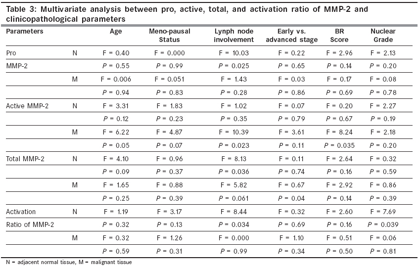
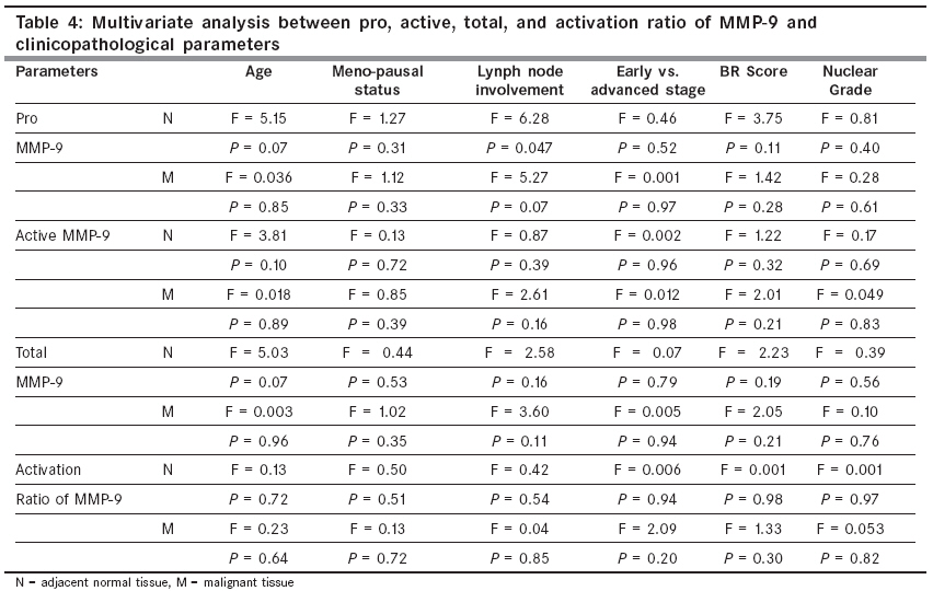
Multivariate analysis of MMP-2 and MMP-9 to evaluate their association with clinicopathological parameters
Multivariate analysis was performed to evaluate the possible correlation of latent, active, and total forms as well as activation ratio of MMP-2 and MMP-9 with clinicopathological features of breast cancer patients. The clinicopathological parameters included age, menopausal status, lymph node involvement, early and advanced stage of the disease, BR score and nuclear grade of the tumor. The correlation of latent, active, total forms, and activation ratio of MMP-2, with clinicopathological features in adjacent normal and malignant breast tissue, is documented in [Table - 3]. The multivariate analysis revealed significant correlation of pro MMP-2, total MMP-2, and the activation ratio of MMP-2 in adjacent normal tissues and lymph node involvement ( P =0.025, P =0.036, P =0.034, respectively). A significant correlation was also observed for active MMP-2, in malignant tissues and lymph node involvement ( P =0.023). The alterations in total MMP-2 levels were significantly associated with the early and advanced stages of the disease in malignant tissues ( P =0.04). Active MMP-2 was significantly associated with the BR score in malignant tissues ( P =0.035). In adjacent normal tissues, variations in the activation ratio of MMP-2 were significantly associated with the nuclear grade of the tumor. [Table - 4] shows the correlation of latent, active, and total forms as well as the activation ratio of MMP-9 with clinicopathological features, in adjacent normal and malignant breast tissues. In the adjacent normal breast tissues, the alterations in pro MMP-9 were significantly associated with lymph node involvement (P =0.047 ) .
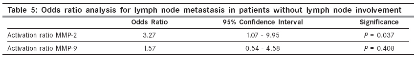
Assessment of risk by odds ratio analysis
Evaluation of risk of development of metastasis in lymph node negative patients is shown in [Table - 5]. The median of activation ratio of MMP-2 and MMP-9 of malignant tissues without lymph node metastasis (0.405 and 0.593, respectively) was considered as the cutoff level. The OR was calculated between, above, and below the cutoff levels of the activation ratio of MMP-2 and MMP-9, of patients with and without lymph node metastasis. The analysis revealed that the activation ratio of MMP-2 above cutoff level was significantly associated with an increased risk of developing lymph node metastasis in node-negative patients (OR=3.27, P =0.037). OR for activation ratio of MMP-9 was also higher, however, it was not statistically significant (OR=1.57, P =0.4).
Discussion
This study evaluates the distribution of MMP-2 and MMP-9 in malignant and adjacent normal breast tissues by zymography. Zymography is a simple, sensitive, quantifiable, cost effective, and functional assay, which identifies MMPs by the degradation of their preferential substrate and by their molecular weight. A distinct separation and determination of different forms of MMPs, that is, the latent and active forms, is the most advantageous feature of this technique.
Using gelatin zymography, we found a total of four different forms of MMP-2 and MMP-9 in malignant and adjacent normal breast tissues. The levels of pro MMP-2 were significantly higher in the adjacent normal tissues, whereas, the active MMP-2, total MMP-2, and activation ratio of MMP-2 levels were significantly higher in malignant breast tissues. Pro MMP-9 levels were comparable between malignant tissues and adjacent normal tissues. Active MMP-9 and activation ratio of MMP-9 were significantly higher in malignant breast tissues as compared to adjacent normal tissues. Paired sample analysis showed that active MMP-2 and MMP-9 levels were 2.60- and 1.29-fold higher, respectively, in malignant breast tissues as compared to adjacent normal tissues. In accordance with the results of the current study, Garbett et al. (1999), found latent MMP-2 and MMP-9 expression in similar proportions of tumor and normal tissues samples, that is, latent MMP-2 in 100% breast tumors, 100% normal breast, and latent MMP-9 in 100% breast tumors, and 93% normal breast tissue specimens. However, the amounts of these enzymes expressed were significantly higher in the tumor tissues than in the corresponding normal tissues. In case of active MMP-2 and MMP-9, the levels were significantly higher in tumor tissues as compared to normal tissues.[10] Remacle et al. (1999), showed the expression of two activated forms of MMP-2. From 84 breast carcinomas they found pro MMP-2 in 12% samples, while its two activated forms (i.e., 62 kDa and 59 kDa) were found in 6% and 40% samples, respectively. In contrast to MMP-2, 52% breast cancer samples contained pro MMP-9, while 10% samples showed the presence of active MMP-9. [7] Liu et al. (2006), also reported significantly higher levels of pro and active MMP-2 and MMP-9 in the tumor tissues than in the corresponding paired adjacent normal tissues. [6] Various studies, by gelatin zymography, showed that active MMP-2 and active MMP-9 levels were higher in malignant tissues as compared to their normal counterparts. [11],[12],[13],[14],[15],[16]
In the current study, higher levels of pro MMP-2 in the adjacent normal tissues were observed, which could be due to a higher induction of MMP-2 in the stromal cells that needed activation for secretion. Furthermore, the absence of a membrane-type MMP-1 (MT MMP-1) and tissue inhibitor of metalloproteinase - 2 (TIMP-2) could hinder the activation of MMP-2. Previously, it has been observed that MMP-2 is synthesized by fibroblasts immediately surrounding the tumor cell clusters and several groups have reported selective protein expression exclusively in the stromal cells adjacent to the areas of tumor cell invasion. [17] It has also been suggested that stromal fibroblasts secrete MMPs, which are stored and activated in carcinoma cells. [18] It is also observed that, MMP-2 and MMP-9 are not produced by the malignant epithelium itself, but rather by the surrounding tumor stroma. In addition to that pro MMP-2 is constitutively expressed by many cell types. [19] The comparable levels of pro MMP-9, between adjacent normal and malignant tissues, in the current study, may be due to the fact that although MMP-9 expression is localized in the tumor stroma, its expression tends to be more focal. [20] In general, the induction of MMPs in the stroma within the tumors and in the adjacent normal tissues represents a direct or indirect host response to the presence of tumor cells.
ROC curve analysis is a more meaningful way to evaluate the discriminatory efficacy of parameters between two groups in the study. It simultaneously provides the sensitivity and specificity of the markers. The ROC curve analysis in the current study shows that pro MMP-2 and active MMP-2 levels can significantly discriminate between malignant and adjacent normal breast tissues. It is reported that the ROC curve for MMP-9 can significantly discriminate between control and head and neck squamous cell carcinoma patients.[21] For the activation ratio of MMP-2 and MMP-9, it is worthwhile to study the activation of MMP-2 and MMP-9, as the latent and active forms of MMP-2 and MMP-9 show elevation with different patterns and sometimes it is difficult to discrete latent and active forms of MMPs. Thus activation ratio is more meaningful way to express gelatinolytic activity of MMP-2 and MMP-9. In the current study, ROC curve for activation ratio of MMP-2 and MMP-9 showed that activation ratio of MMP-2 could significantly discriminate between patients with lymph node metastasis and without metastasis. In accordance with the results of the current study, higher activation ratio of MMP-2 and MMP-9 are documented by several studies.[16],[22],[23],[24]
In the present investigation, multivariate analysis revealed a significant correlation between pro MMP-2, total MMP-2, and activation ratio of MMP-2 in adjacent normal tissues and lymph node involvement. A significant correlation was also observed between active MMP-2 in malignant tissues and lymph node involvement. The alterations in total MMP-2 levels were significantly associated with the early and advanced stages of the disease in malignant tissues. Active MMP-2 was significantly associated with the BR score in the malignant tissue. In adjacent normal tissues, variations in the activation ratio of MMP-2 were significantly associated with the nuclear grade of the tumors. The alterations in pro MMP-9 were significantly associated with lymph node involvement in the adjacent normal breast tissues. Liu et al. (2006), reported significantly increased MMP-2 levels in patients with metastasis and also showed that the MMP-2 level was significantly different between tumor grades and were positively correlated with the tumor size in the tumor tissue samples. [6]
Currently, the nodal status is the strongest prognostic factor in breast cancer and systemic adjuvant treatment is offered to as many as 90% of the breast cancer patients. This implies that a number of patients are overtreated, especially in the group of lymph node negative patients, among whom ~60-70% are cured by surgery alone. [25] In breast carcinoma, MMP-2 expression in the tumor tissue has been associated with unfavorable prognosis in both node negative and node positive cases. [5],[26],[27],[28] Daidone et al. (1991), have demonstrated a correlation between the expression of MMP-2 immunoreactive protein and local recurrence in node-negative patients. [29] Li et al. (2004), have reported that the coexpression of MMP-2 and MMP-9 has significant prognostic value in node-negative patients, for predicting relapse-free survival. [30] The significant association of MMP-2 and MMP-9 with lymph node involvement in breast cancer, in the present study, led us to analyze the risk of lymph node metastasis in patients without lymph node involvement. The higher odds ratio for the activation ratio of MMP-2 in lymph node negative tissues suggested increased risk of metastasis and therefore it could be used to monitor patients without lymph node involvement. These patients should be followed up closely to find out the regional metastasis. Earlier report from our laboratory also observed similar results in oral cancer. [16] The significant correlation of activation ratio of MMP-2 with lymph node metastasis, in the current study, suggested that MMP-2 could be useful as the major drug target in the treatment of breast cancer.
Conclusion
To the best of our knowledge this is the first Indian study in which gelatin zymography was used to resolve the activated form from the latent proenzyme of gelatinases (MMP-2 and MMP-9), in breast cancer. Our results on the expression of active MMP-2 and MMP-9 endorse the significance of these proteolytic enzymes in tumor invasion and metastasis in breast cancer. The higher activation ratio of MMP-2 in breast cancer patients without lymph node involvement poses a higher risk of development of regional lymph node metastasis in these patients, which may be helpful for planning the systemic adjuvant treatment for lymph node-negative breast cancer patients. The data also suggests that inhibition of MMP-2 activation would be an interesting approach toward a biological therapy for breast cancer. Thus, in addition to the significant clinical usefulness of the data, the present investigation could be an important contribution in developing a targeted therapy for breast cancer.
References
| 1. | Parkin DM, Bray F, Pisani P. Global Cancer Statistics, 2002. CA Cancer J Clin 2005;55:74-108. Back to cited text no. 1 [PUBMED] [FULLTEXT] |
| 2. | Hanahan D, Weinberg RA.The hallmarks of cancer. Cell 2000;100:57-70. Back to cited text no. 2 [PUBMED] [FULLTEXT] |
| 3. | Egeblad M, Werb Z. New functions for the matrix metalloproteinases in cancer progression. Nat Rev Cancer 2002;2:161-74. Back to cited text no. 3 [PUBMED] |
| 4. | Strenlicht MD, Werb Z. How matrix metalloproteinases regulate cell behavior. Ann Rev cell Dev Biol 2001;17:463-516. Back to cited text no. 4 |
| 5. | Talvensaari-Mattila A, Paakko P, Hoyhtya M, Blanco-Sequeiros G, Turpeenniemi-Hujanen T. Matrix metalloproteinases-2 immunoreactive protein: A marker of aggressiveness in breast carcinoma. Cancer 1998;83:1153-62. Back to cited text no. 5 |
| 6. | Liu SC, Yang SF, Yeh KT, Yeh CM, Chiou HL, Lee CY, et al . Relationship between the level of matrix metalloproteinase-2 and tumor size of breast cancer. Clin Chim Acta 2006;371:92-6. Back to cited text no. 6 [PUBMED] [FULLTEXT] |
| 7. | Ramacle AG, Noel A, Duggan C, McDermott E, O'Higgins N, Foidart JM, et al . Assay of matrix metalloproteinases types 1,2,3 and 9 in breast cancer. Br J Cancer 1998;77:926-31. Back to cited text no. 7 |
| 8. | Lowry OH, Rosebrough NJ, Farr AL, Randali RJ. Protein measurement with the folin phenol reagent. J Biol Chem 1951;193:265-75. Back to cited text no. 8 |
| 9. | Lorenzo JA, Pilbeam CC, Kalinowski JF, Hibbs MS. Production of both 92 and 72 kDa gelatinase by bone cells. Matrix 1992;12:282-90. Back to cited text no. 9 [PUBMED] |
| 10. | Garbett EA, Reed MW, Brown NJ. Proteolysis in human breast and colorectal cancer. Br J Cancer 1999;81:287-93. Back to cited text no. 10 [PUBMED] [FULLTEXT] |
| 11. | Hrabec E, Strek M, Nowak D, Greger J, Suwalski M, Hrabec Z. Activity of type IV collagenases (MMP-2 and MMP-9) in primary pulmonary carcinomas: a quantitative analysis. J Cancer Res Clin Oncol 2002;128:197-204. Back to cited text no. 11 [PUBMED] [FULLTEXT] |
| 12. | Baker EA, Leaper DJ. The plasminogen activator and matrix metalloproteinase systems in colorectal cancer: Relationship to tumour pathology. Eur J Cancer 2003;39:981-8. Back to cited text no. 12 [PUBMED] [FULLTEXT] |
| 13. | Sier CF, Casetta G, Verheijen JH, Tizzani A, Agape V, Kos J, et al . Enhanced gelatinase urinary activity (Matrix metalloproteinases 2 and 9) are associated with early stage bladder carcinoma: A comparison with clinically used tumor markers. Clin Cancer Res 2000;7:445-7. Back to cited text no. 13 |
| 14. | Schutz A, Schneidenbach D, Aust G, Tannapfel A, Steinert M, Wittekind C. Differential expression and activity status of MMP-1, MMP-2 and MMP-9 in tumor and stromal cells of squamous cells carcinoma of the lung. Tumor Biol 2002;23:179-84. Back to cited text no. 14 |
| 15. | Hong SD, Hong SP, Lee JI, Lim CY. Expression of matrix metalloproteinase-2 and -9 in oral squamous cell carcinoma with regard to the metastatic potential. Oral Oncol 2000;36: 207-13. Back to cited text no. 15 [PUBMED] [FULLTEXT] |
| 16. | Patel BP, Shah PM, Rawal UM, Desai AA, Shah SV, Rawal RM, et al. Activation of MMP-2 and MMP-9 in patients with oral squamous cell carcinoma. J Sur Oncol 2005;90:81-8. Back to cited text no. 16 |
| 17. | Polette M, Clavel C, Cockett M, Girod de Bentzmann S, Murphy G, Birembaut P. Detection and localization of mRNAs encoding matrix metalloproteinases and their tissue inhibitors in breast pathology. Invasion Metastasis 1993;13:31-7. Back to cited text no. 17 |
| 18. | Polette M, Gilbert N, Stas I, Nawrocki B, Noel A, Remacle A, et al . Gelitinase A expression and localization in human breast cancers. An In situ hybridization study and immunohistochemical detection using confocal microscopy. Virchow Arch 1994;424:641-5. Back to cited text no. 18 |
| 19. | Brown PD, Levy AT, Margulies IM, Liotta LA, Stetler-Stevenson WG. Independent expression and cellular processing of Mr72000 type IV collagenase and interstitial collagenase in human tumourigenic cell lines. Cancer Res 1990;50:6184-91. Back to cited text no. 19 [PUBMED] [FULLTEXT] |
| 20. | Heppner KJ, Matrisian LM, Jensen RA, Rodgers WH. Expression of most matrix metalloproteinases family members in breast cancer represents a tumour induced host response. Am J Pathol 1996;149:273-82. Back to cited text no. 20 [PUBMED] [FULLTEXT] |
| 21. | Ranuncolo SM, Matos E, Loria D, Vilensky M, Rojo R, Bal de Kier Joffe E, et al . Circulating 92- Kilodalton matrix metalloproteinase (MMP-9) activity is enhanced in the euglobulin plasma fraction of head and neck squamous cell carcinoma. Cancer 2002;94: 1483-91. Back to cited text no. 21 |
| 22. | Schmidt M, Polednik C, Hoppe F. Proteolytic patterns of head and neck squamous cell carcinoma. Eur Arch Otorhinolaryngol 1999;256:346-50. Back to cited text no. 22 [PUBMED] [FULLTEXT] |
| 23. | Koshiba T, Hosotani R, Wada M, Miyamoto Y, Fujimoto K, Lee JU, et al . Involvement of matrix metalloproteinase-2 activity in invasion and metastasis of pancreatic carcinoma. Cancer 1998;82:642-50. Back to cited text no. 23 [PUBMED] [FULLTEXT] |
| 24. | Takashi M, Oka N, Naroda T, Nishitami MA, Kanda K, Kanayama HO, et al . Prognostic significance of matrix metalloproteinases-2 activation ratio in renal cell carcinoma. Int J Urol 2002;10:531-8. Back to cited text no. 24 |
| 25. | Schrohl AS, Holten Andersen MN, Peters HA, Look MP, Meijer-van Gelder ME, Klijn JG, et al . Tumor tissue levels of Tissue inhibitor of metalloproteinase-1 as a prognostic marker in primary breast cancer. Clin Cancer Res 2004;10:2289-98. Back to cited text no. 25 |
| 26. | Talvensari-Mattila A, Pakko P, Turpeenniemi-Hujanen T. MMP-2 positivity and age less than 40 years increases the risk for recurrence in premenopausal patients with node positive breast carcinoma. Breast Cancer Res Treat 1999;58:287-93. Back to cited text no. 26 |
| 27. | Talvensari-Mattila A, Pakko P, Blanco-Sequeiros G, Turpeenniemi-Hujanen T. Matrix metalloproteinases-2 is associated with the risk for a relapse in postmenopausal patients with node positive breast carcinoma treated with antiestrogen adjuvant therapy. Breast Cancer Res Treat 2001;65:55-61. Back to cited text no. 27 |
| 28. | Hirvonen R, Talvensari-Mattila A, Pakko P, Turpeenniemi-Hujanen T. Matrix metalloproteinases-2 (MMP-2) in T (1-2) N0 breast carcinoma. Breast Cancer Res Treat 2003;77:85-91. Back to cited text no. 28 |
| 29. | Daidone MG, Silvestrini R, D'errico A, Di Fronzo G, Benini E, Mancini AM, et al . Laminin receptors, collagenase IV and prognosis in node-negative breast cancers. Int J Cancer 1991;48: 529-32. Back to cited text no. 29 [PUBMED] |
| 30. | Li HC, Cao DC, Liu Y, Hou YF, Wu J, Lu JS, et al . Prognostic value of matrix metalloproteinases (MMP-2 and MMP-9) in patients with lymph node-negative breast carcinoma. Breast Cancer Res Treat 2004;88:75-85. Back to cited text no. 30 [PUBMED] [FULLTEXT] |
Copyright 2009 - Indian Journal of Cancer
The following images related to this document are available:
Photo images
[cn09043f1.jpg]
[cn09043f3.jpg]
[cn09043t2.jpg]
[cn09043t4.jpg]
[cn09043t5.jpg]
[cn09043f2.jpg]
[cn09043f4.jpg]
[cn09043t3.jpg]
[cn09043t1.jpg]
|
