 |
| About Bioline | All Journals | Testimonials | Membership | News |
|
||||||
|
||||||
Indian Journal of Cancer, Vol. 46, No. 4, October-December, 2009, pp. 297-302 Original Article Lipid peroxidation and antioxidants in different stages of cervical cancer: Prognostic significance Srivastava S, Natu SM, Gupta A, Pal KA, Singh U, Agarwal GG, Singh Uma, Goel MM, Srivastava AN Department of Pathology, C. S. M. Medical University, Lucknow Code Number: cn09071 PMID: 19749459 Abstract Background: Free radical Injury is associated with cancer, but how the extent of oxidative stress correlates with the FIGO (International Federation of Gynecology and Obstetrics) stage in Carcinoma Cervix (Ca Cx), and its significance as a prognostic marker, is not clear and needs an in-depth study.Aim: To correlate the blood levels of Lipid Peroxidation (LPO), Reduced Glutathione (GSH), Superoxide Dismutase (SOD), and Vitamin A and E levels with the clinical stage in Ca Cx. Settings and Design: This is a Prospective Case Control Study. Materials and Methods: LPO, SOD, reduced GSH were estimated by Bio Chemical Assays and Vitamins by High Performance Liquid Chromatography (HPLC). Statistical Analysis: The cases and controls were compared using One Way ANOVA and different stages over different time periods were individually compared by Repeated Measure Analysis of Variance. Results: The results indicated a statistically significant increase of LPO vis-a-vis the FIGO stage of Ca Cx and control, while the antioxidant status as depicted by GSH and SOD decreased. Vitamin A and E levels were significantly lower in cancer cases as compared to the control. Conclusion: Increased LPO and reduced antioxidant levels may be taken as associated predictive markers, thus suggesting that Ca Cx cases should get nutritive supplements to contain the blood LPO level and maintain a positive balance of antioxidants for a better outcome in terms of delayed recurrence and better Quality of Life (QOL). Keywords: Antioxidants, cervical cancer, free radicals, prognosis Introduction Cervical cancer is the most prevalent genital tract cancer in the world, including India. [1],[2] It is a multifactorial disease process and several risk factors include, early age intercourse, multiple sex partners, low socioeconomic status, and Human papillomavirus (HPV) infection. [1],[2] Chronic inflammation and infection over a prolonged period of time have been recognized as major risk factor for disease initiation. [3] Carcinoma in situ is but a phase leading to frank cancer. [4] Evidence has indicated that reactive oxygen species (ROS) are involved in the initiation and progression of carcinogenesis. This may be due to the damage caused to the tumor suppressor genes or immunological defenses in our body. [5] Superoxide and hydroxyl radicals are oxygen-free radicals, involved in producing oxidative stress. This oxidative stress can be associated with other factors which may lead to various neoplastic transformations. [6] Deleterious effects of these oxidants are counteracted by antioxidants such as Superoxide dismutase (SOD), Glutathione peroxidase (GPX), and reduced glutathione (GSH). [7] In addition to the body′s defense mechanism there are vitamins that provide the body with the much-needed immunity and a mechanism of self-defense to fight against various pathogens. Studies indicate that the level of these antioxidants in the body decrease in cases of carcinogenesis. The levels of vitamin E were found to vary in a study of cervical carcinogenesis. [8] Very few studies have given importance to different stages of cervical cancer as per FIGO staging/grading and degree of free radical damage correlations. The present study was therefore planned to study free radical injury levels and some antioxidants in the four stages of cervical cancer cases, before and after treatment. Materials and Methods Subjects Subjects included in this study comprised women attending the Out door Patients Department (OPD) of Obstetrics and Gynecology of our institution. Of the 356 registered patients suspected for cervical pathology; pap smears of all the 356 cases were collected and after detection of cancer in the Pap smear reports and further confirmation by histopathological reports, (taken as Gold Standard) the cases were enrolled in the study. Any patient with a history of prior treatment for cancer or any previous associations with a chronic debilitating disease like HIV, TB, and so on were excluded from the study. Thus 95 cases were finally enrolled for this study. All these patients received radiation treatment, which was as follows:
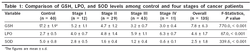
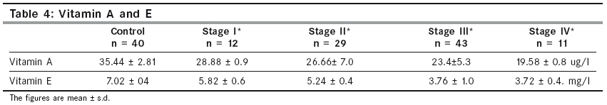
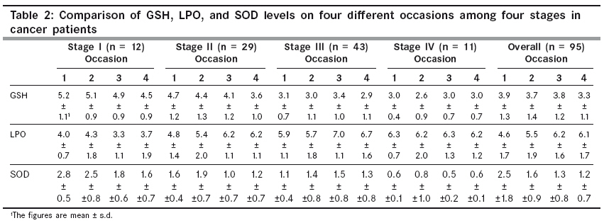
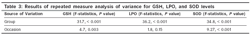
Venous blood samples of all these patients were collected in heparinzed tubes for separation of plasma and lysate, for bio-chemical assays. Five milliliters of venous blood samples were collected on Day ′0′, after 6-8 weeks, 6-8 months, and 1 year after starting the treatment. Serum, plasma, and haemolysate were separated from these blood samples. For plasma and haemolysate preparation blood was taken in the EDTA vials. Estimation of lipid peroxide levels (LPO) Blood plasma of 0.2 ml was mixed with 1 ml of 20% acetic acid; subsequently 0.2 ml of 8% SDS was added to the above mixture and pH was adjusted to 4. Following that, 1.5 ml of 0.8% TBA and 1.1 ml of distilled water was added. This reaction mixture was incubated in a boiling water bath for 1 hour. After cooling, 3 ml of n-butanol was mixed, and then centrifuged at 10,000 g for 15 minutes. A clear butanol fraction thus obtained was used for measuring the absorbance at 532 nm. [9] Enzyme purification for reduced glutathione and superoxide dismutase Haemolysate of 0.2 ml was mixed with 0.8 ml chilled water, 0.5 ml ethanol, and 0.25 ml chloroform. The mixture was stirred and kept for 15 minutes at 4˚C. It was then centrifuged at 3000 rpm for 15 minutes. The precipitate was discarded and the supernatant used for estimation of GSH and SOD. According to Tsuchihashi M (1923), [10] the precipitation step helps in the removal of hemoglobin from haemolysate. [11],[12],[13],[14] Estimation of reduced glutathione (GSH) One milliliter of the supernatant after ethanol and chloroform extraction as above was mixed with 1 ml DTNB reagent just before measuring the absorbance of the sample at 412 nm. GSH solution of known concentration was similarly processed to prepare a standard curve. The amount of GSH in the sample was determined from the standard curve. [15] Estimation of superoxide dismutase (SOD) The supernatant from the above extraction step was divided into two portions, Experimental and Reference. To the experimental test tube we added 0.3 ml Nitroblue tetrazolium (NBT), 0.2 ml Phenazine Metho Sulphate (PMS) and 0.2 ml pyrophosphate buffer, 1 ml D.D.W, and 2 ml enzyme for analysis. In the reference test tube, everything was added along with NADH except the enzyme.The reaction was run for 90 seconds at 37°C with constant stirring. The reaction was stopped by adding 1 ml acetic acid. After 10 minutes, the enzyme was added in the reference test tube and optical density (OD) was read at 560 nm. Blank included NBT, PMS Buffer, and Triple Distilled Water. [16] Estimation of protein Haemolysate was used for protein estimation. Haemolysate of 0.1 ml was taken and to this was added 0.9 ml of triple distilled water. Further, 1 ml 10% trichloroacetic acid was added and then kept at 4°C for 4 hours. After centrifugation at 4000 rpm for 15 minutes, the supernatant was discarded and the precipitate was dissolved in 2 ml 0.1 N NaOH. For protein estimation 0.1 ml of the sample was taken as protein. To this was added alkaline Cu reagent and incubated for 20 minutes at 37°C, then Folin′s reagent was added and incubated at 37°C for 15 minutes. Bovine Serum Albumin (BSA) was used as a standard and OD was read at 660 nm. [17] Estimation of vitamins Vitamin A and E were measured by high performance liquid chromatography (HPLC) as per the modified method of Omu et al . [18] Briefly α -tocopherol acetate and retinol acetate were pipetted into an Eppendorf tube.To this, blood serum was added and vortex mixed; hexane extract of vitamins A and E was aspirated out into a glass tube, dried under nitrogen stream, and dissolved into methanol. Finally this preparation was injected into a HPLC, fitted with reverse phase of C 18 stainless steel column.The vitamins were eluted with methanol at a flow rate of 1.5 ml /min for 15 minutes.The peak heights and curve areas of vitamin A and E and their acetates were measured to calculate the amount of these vitamins in the blood with an ultraviolet detector with 292 nm filters. The levels of vitamin A and E were not planned for estimation at different occasions due to financial constraints of the study. Statistical Analysis At the time of the recruitment (occasion 1), five groups of subjects (control and four stages of cancer patients) were available, so one-way analysis of variance (ANOVA) was performed to test the equality of mean GSH, LPO, and SOD levels. The longitudinal observations were measured on four different occasions for four stages of cancer patients. To take into account the correlated behavior of longitudinal observations, the repeated measure analysis of variance was performed to compare the mean GSH, LPO, and SOD levels among the four groups as well as over four different occasions. The P-value of < 0.05 was taken to be significantly different. For the purpose of analysis we have defined the four time periods as Occasion 1: day ′0′, Occasion 2: as 6-8 weeks, Occasion 3: as 6-8 months and Occasion 4: as1 year. Results The results of LPO, GSH, and SOD in different stages of cervical cancer and that of healthy controls at day ′0′ are shown in [Table - 1]. The results revealed increase in levels of LPO with higher stage of cervical cancer and the lowest levels were seen in the control group. GSH and SOD in control samples showed higher values, which decreased significantly in the higher cancer stages. The one-way ANOVA showed that the biochemical levels in [Table - 1] and the level of vitamins in [Table - 4] in all the four stages of cervical cancer patients as well as the control were significantly different ( P < 0.001). The differences were most significant for GSH (F-value = 770.0) followed by SOD (F-value = 339.6) and LPO (F-value = 67.0). The patients were followed on four different occasions (day ′0′, 6-8 weeks, 6-8 months, and 1 year).The measurements were not paralleled with that of control for reasons explained earlier. The comparisons of four different stages for four different occasions are shown in [Table - 2]. The mean GSH values showed a decreasing pattern for the four cancer stages as well as on the four occasions. The overall status was decreasing over the four time periods. The mean SOD values as shown in [Table - 2] decreased with stages and the decrease was also consistent for the different occasions. The overall values showed similar results as those for GSH. The mean LPO levels showed an increasing pattern over the four stages on occasions 1 and 2. The same increasing pattern was observed over the four occasions for stage I, stage II, and stage III. However, in stage IV the LPO levels exhibited an inconsistent pattern on occasion 4. The results of repeated measures of variance analysis with different time points as "within subject factor" and different stages of cancer as "between factors" are depicted in [Table - 3]. All the three biochemical parameters were significantly different for the four stages of cancer ( P < 0.001). However, when the comparison was made on different occasions GSH and SOD levels were significantly different with P =0. 003 and P < 0.001, respectively. The LPO level on different occasions was not significant ( P = 0.15). These results are in coherence with the results of [Table - 2], as discussed above. Vitamin levels were significant, P < 0.001 when the values were compared with controls on day ′0′. Discussion Cervical cancer is the most common cancer in women in developing countries. One of the main causes of this high prevalence is the lack of awareness in women for its early detection and management. A number of risk factors have been associated with cervical pre cancer and cancer. An extensive search over the past several years has suggested the role of free radicals in a number of diseases including carcinogenesis. [19] Although the body′s own defense mechanism plays a crucial role to control the levels of these free radicals, the levels of antioxidants that counterbalance these oxidative radicals get impaired themselves. The present study was planned to detect the levels of LPO, SOD, GSH, and Vitamin A and E in cases of uterine cervical cancer in different FIGO stages I, II, III, and IV. Free radical detection was done in controls and patients at day ′0′, that is, before the start of the treatment. Later on, the measurements of LPO, GSH, and SOD were done after 6-8 weeks, after 6-8 months, and after one year of starting the treatment, in the same set of patients. In the present study the values of LPO demonstrated higher levels in stages I to IV as compared to control and the differences were statistically significant. However repeated measure analysis of variance values at different intervals had an inconsistent pattern and there was statistically no significant difference at day ′0′, 6-8 weeks, 6-8 months, or a 1 year level.It appears that a saturation plateau was reached in Stage IV disease as far as the plasma circulating level of lipid peroxide was concerned. Lipid Peroxidation is the oxidative conversion of polyunsaturated fatty acids to MDA (malondialdehyde); which is cytotoxic and acts as a tumor promoter and a co-carcinogenic agent. [3],[4],[5],[6],[7],[8],[9],[10],[11],[12],[13],[14],[15],[16],[17],[18],[19],[20] Damage caused by LPO impairs the functioning of the biological membrane and the continued damage leads to loss of membrane integrity. [21] Beevi et al . [22] observed increased plasma as well as erythrocyte MDA in patients with cervical cancer, although there was no significant variation in lipid peroxides according to the stage of the tumor in their study. Manju et al. [23] and Kim et al. , [24] have similarly demonstrated the involvement of rising LPO and compromised antioxidant levels. Ahmed et al [25] in their study demonstrated an overall progressive impaired status of antioxidants in Ca Cx. Manoharan et al. [26] have demonstrated enhanced erythrocyte LPO and impaired antioxidant enzyme activities, suggesting avenues for exploring damage to the red cell membrane structure and function in cervical cancer. An analysis of reduced GSH in comparison to control showed consistent low values. The patients demonstrated significant decrease of reduced GSH levels from day ′0′ to 6-8 weeks, 6-8 months and finally up to 1 year. Pejic et al [7] have shown in their study a similar observation about high levels of lipid peroxides and markedly reduced levels in various gynecological conditions including cancer and have shown significant decrease of GSH level as well. Depleted levels of GSH in cervical cancer cases may be related to the reduction in activity of glucose-6-phosphate dehydrogenase in cervical cancer patients. [16] Nicotinamide Adenosine Dinucleotide Phosphate (NADPH) produced by the catalyst glucose-6-phosphate dehydrogenase plays an important role in the reduction of oxidized GSH to reduced GSH. [27] This study revealed a very consistent reduction in the levels of SOD as compared to the control in all the stages at the time of patient recruitment. At different occasions, that is, at day ′0′, 6-8 weeks, 6-8 months and 1 year statistically significant lowering of SOD levels were seen. Vitamin A is a known antioxidant which is especially responsible for healthy development and upkeep of the epithelium. Deficiency of vitamin A exposes the patient to further affects of oxidative stress, inhibiting any cell repair and return to normalcy. The levels of vitamin A in our studies showed a statistically significant diminution between various stages of cervical cancer from Stage I to Stage IV. This shall be an interesting idea to differentially investigate antioxidants and vitamin A in various stages of cervical cancer at day ′0′ levels and compare it with levels at various intervals in groups of patients who received vitamin A and in those who did not receive it. This may possibly also reveal differences in the levels of antioxidant status in terms of SOD, GSH, and so on. Manju et al., [23] demonstrated a significantly diminished value for vitamin E level along with reduction in other antioxidants such as GSH, GPX, and SOD, and attributed this reduction to the increased utilization in scavenging lipid peroxides as well as their sequestration by tumor cells. Their case control study was limited to a group of cervical cancer cases en-block (without any stagewise differentiation), whereas, in the present study the levels of vitamin E were studied in different stages of cervical cancer and showed a consistent difference among stages I, II, III, and IV, when mutually compared..Ahmed et al (1999) have shown that oxygen free radical levels are raised in cervical dysplasia and cervical cancer as compared to controls. [25] There are only a few studies showing FIGO stagewise measurements of vitamin E / vitamin C and other antioxidants in cervical cancer cases to correlate these findings. [23],[24],[25],[26],[27],[28] The present study fills this gap to some extent and documents FIGO stagewise levels in SOD, GSH, and vitamin A and E. The higher the FIGO stage the lower the level of antioxidants in blood. Other factors remaining same, the most important factor that can influence the levels of antioxidants in blood is the poor socioeconomic status of the patients, responsible for deficient nutrient intake. In the present study > 95% of patients was of lower middle class, thus eliminating this confounder. In summary we can say that LPO is high in cases of cervical cancer as compared to controls when measured at day ′0′ and is statistically significant, although on different later occasions the levels become statistically insignificant. The levels of SOD and reduced GSH, which are already low on day ′0′, further dip down significantly at higher stages of cancer and this decrease is statistically significant. The levels of vitamin A and vitamin E show significant lower values in cancer patients and healthy controls on day ′0′ and the level dips as the stage of cancer gets higher. This study gives the thought that carcinoma cervix cases, in addition to regular chemo-radiotherapy, may be given antioxidants to contain LPO levels, and to maintain a positive balance of antioxidants for better outcome in terms of delayed recurrence and better quality of life. Further studies comprising larger series of patients are suggested to corroborate these findings. Acknowledgments The authors thank the Himalayan Drug Company, Bangalore, for partially financing this study. References
Copyright 2009 - Indian Journal of Cancer The following images related to this document are available:Photo images[cn09071t4.jpg] [cn09071t1.jpg] [cn09071t3.jpg] [cn09071t2.jpg] |
| |||||||||
