 |
| About Bioline | All Journals | Testimonials | Membership | News |
|
||||||
|
||||||
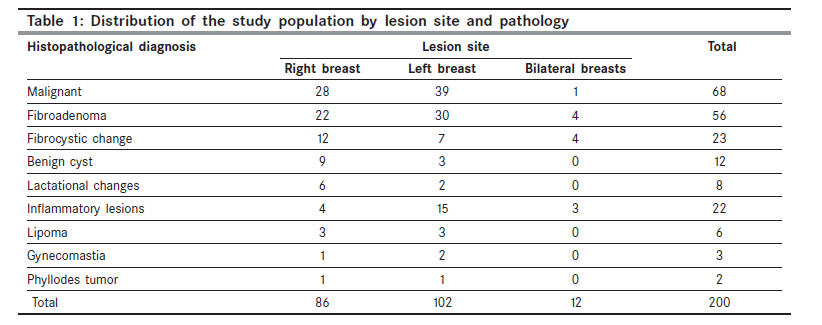
Indian Journal of Cancer, Vol. 47, No. 1, January-March, 2010, pp. 23-26 Original Article Frequency of breast cancer among Sudanese patients with breast palpable lumps Ahmed HG, Ali AS, Almobarak AO1 Department of Histopathology and Cytology, Faculty of Medical laboratory Sciences, University of Khartoum, Khartoum, Sudan; 1 Alrebat University, Khartoum, Sudan Code Number: cn10006 PMID: 20071785 DOI: 10.4103/0019-509X.58854 Abstract Background: Breast cancer mortality is high in Sudan and most patients are detected at later stages of the disease due to the lack of awareness and absence of screening programs. This study aimed to determine the pattern and frequency of breast cancer among patients presenting with palpable breast lumps within one year duration. Settings and Design: As a part of the continuous development in cancer management, this descriptive longitudinal study was conducted in Khartoum, Sudan. Methods and Materials: We obtained information (patient's personal data) and Fine-Needle Aspiration (FNA) materials, for occurrence of 200 breast lesions in patients. Statistical Analysis Used: Data were analyzed by using a computer SPSS program. Results: The diagnoses of the 200 breast lesions were as follows: 68 (34%) were malignant, 56 cases (28%) were fibroadenoma, 23 cases (11.5%) were fibrocystic change, 22 cases (11%) were inflammatory lesions (including mastitis and abscess formation), 12 cases (6%) were benign cysts and the remaining 19 patients (9.5%) were with lactation changes (8 cases), lipoma (6 cases), gynecomastia (3 cases) and phyllodes tumor (2 cases). Regarding gender, only 6 patients (0.03%) were males of whom 3 (50%) were diagnosed with gynecomastia. Conclusions: The frequency of advanced breast cancer among patients with breast lesions is high, in this subset of patients, which signals the urgency for implementation of breast screening programs.Keywords: Breast cancer, sudan, women, fine-needle aspiration Introduction Breast cancer is a public health problem worldwide; therefore, it is critical that efforts in prevention and early diagnosis of breast cancer are implemented everywhere. One of the main problems concerning breast cancer relates to the lack of patients awareness about the disease. Limitations in implementing breast self-examination and mammography screening programs are the other important issues. Overall survival and mortality due to this disease are influenced strongly by the stage of the disease at diagnosis. About 54% of the women are diagnosed in stage II, while only 16% are diagnosed in stage I. [1] Breast awareness is a part of general body awareness. Learning how your breasts feel at different times will help you to know what is normal for you. The introduction of mammographic screening has led to an increased detection of breast cancers, at early stages. [2] Fine-needle aspiration cytology (FNAC) is part of the triple assessment for the diagnosis of breast lesions. It is an established, highly accurate method for diagnosing breast cancer and has given rise to a reduction in the number of excision biopsies for benign breast disease. [3] This study aimed at identifying the type of breast lesions in one cytodiagnosis center in Khartoum, Sudan, and to determine the frequency of breast cancer among these patients. Materials and Methods The frequency of breast cancer was estimated in 200 patients with palpable breast lumps who were attended in a diagnostic center in Khartoum. This study included all patients with breast lesions who were attended during a period of one year. FNAC was introduced as a preoperative diagnostic tool in the investigations of palpable breast lumps. A total of 200 patients with breast masses were selected depending on the clinical discovery of a palpable mass in the breast. A questionnaire to obtain essential data about the patient was used, and also each patient was asked to sign a written ethical consent. A total of 200 breast lesions underwent FNAC, of which 131 were subsequently confirmed by histopathological examinations. All malignant and clinically suspicious lesions were confirmed by histopathological examinations. Statistical analysis: Data were analyzed by using a computer SPSS program. Pearson χ2 test was used for statistical analyses. Results FNA materials and/or biopsies were obtained from 200 patients (194 females and 6 males) with breast palpable lumps. The ages of the study subjects ranged from 15 to 85 years, with a mean age of 37 years. The diagnoses of the 200 breast lesions were as follows: 68 (34%) were malignant, 56 cases (28%) were fibroadenoma, 23 cases (11.5%) were fibrocystic change, 22 cases (11%) were Inflammatory lesions (including mastitis and abscess formation), 12 cases (6%) were benign cysts, and the remaining 19 patients (9.5%) were with lactation changes (8 cases), lipoma (6 cases), gynecomastia (3 cases) and phyllodes tumor (2 cases) as shown in [Table - 1]. Of the 68 malignant cases, 43 (63%) were detected with T4, 16 (23.5%) with T2, 7 (10.2%) were detected with T3 and only 2 (2.9%) were detected with T1. The primary lesion sites were found as follows: left side (102 cases, 51%), right side (86 cases, 43%) and bilateral (12 cases, 6%). Moreover, 57.4% of the malignant lesions and 53.7% of the fibroadenoma lesions were observed at the left side; similarly, 151 (75.5%) of the lesions were found as mobile lumps, and the remaining 49 (25%) were fixed of which 41 (93%) were found to be malignant. Furthermore, of the 200 patients, only 40 (20%) were found with nipple discharges, among whom 16 (40%) were patients with cancer. Concerning the distribution of the study population by age it was apparent that the risk of cancer increased with increase in age, as about 100%, 75%, 71.4% and 85% of the patients with cancer were detected in the age ranges 85-94, 75-84 and 65-74, respectively (P < 0.0001). With regard to the distributions of the study populations by demographical situations the majority of malignant cases were found among Gaaleen tribe followed by Shaggy, Nubein and Baniamer, constituting 15 (22.7%), 12 (18.2%), 10 (15.2%) and 8 (12.1%) correspondingly. According to the residence, most of the patients were from Khartoum state followed by East, West and North representing 77%, 13%, 7% and 3%, respectively. Furthermore, 131 of the patients were married among whom 59 (89.4%) were having malignant changes; conversely, 69 were patients unmarried among whom fibroadenoma was detected in 45 (65.2%). Discussion Cancer continues to be the most common health problem that plagues our civilization, and the current data ensures the increasing importance of the subject. The escalating knowledge in all areas of medicine has made a significant impact on the field of pathology, which is crucial to our understanding of the cause, the course and the management of disease processes. Incidence of breast cancer exceeds all female cancers with high mortality rates worldwide. [4],[5] Female breast cancer is by far the leading cancer in the Sudan. It accounts for 16.12% of all cancers (30.24% of all female cancers in 2006). A vast majority of these cancers were from Khartoum, with 43.7% in 2006, as reported by Radio Isotope Centre Khartoum (RICK), [6] which is the prime center that has prompted these services in Sudan. Sudan is experiencing a rapidly increasing cancer epidemic that carries many challenges that are distinctive of developing countries. These include a high incidence of advanced, complicated stage of the disease at presentation, and a high frequency of cancer that is related to a number of risk factors that are required for stabilization. Of the 68 cancer patients, 80% were detected with an advanced stage of breast cancer. This supplements the evidence that in low-resource countries, most patients present with advance stages of the disease. Therefore, the burden of cancer in developing countries is growing and threatens to exact heavy morbidity and mortality. [7] It is impossible to know whether a breast lump is cancerous without performing imaging examinations and/or a biopsy and/or FNA. However, if the lump in breast is firm, hard and fixed it is more likely to be cancerous. This might be our explanation for the finding of 93% of the malignant lesions in the present series as fixed lumps. Nipple discharge is a common problem of the breast that has been reported in 10%-15% of women with benign breast disease and in 2.5%-3% of women with breast cancer. Nipple discharge should be of concern when a woman reports it as unilateral and spontaneous. [8] However, our findings revealed nipple discharge in 18% of women with benign lesions and in 23.5% of women with breast cancer. These findings supported the fact that both malignant and benign lesions may produce nipple discharge. If the secretion appears clear or milky, yellow or green, cancer is very unlikely. If the discharge is red or red-brown, suggesting that it contains fresh blood or old blood, it might be due to cancer. Nipple discharge alone is not usually a sign of breast cancer. [9],[10] However, about 100%, 75%, 71.4% and 85% of the cancer patients were detected at age ranges 85-94, 75-84 and 65-74, respectively. These results indicate that the risk for breast cancer increases with the increase in age. These findings were widely advocated in the literature by numerous studies. The incidence of the cancer of the breast increases through a woman's life time. About 77% incidence of cancer occur in women over 50 years of age. The average age at diagnosis is 64 years. [11] The prevalence of fibroadenomas is approximately 8%-10% in women older than 40 years. Fibroadenomas are the second most common solid tumor after breast cancer and the most common benign tumor in women. In women younger than 30 years, fibroadenoma is the most commonly diagnosed breast tumor. In the current study, fibroadenomas represent 42.4% of all identified benign conditions and most cases were detected among non-married patients. It was previously reported that fibroadenoma can occur in all age groups, but it is especially seen in young women from 20 to 35 years of age. [12] A majority of these lesions were observed on the left side (102 of cases), 86 patients were having the lesion on the right side and only 12 were having bilateral lesions. Of the 68 patients with cancer, 39 were detected with left lesions; hence, 28 were on the right side. We could not find anything in the literature, particularly with regard to occurrence of breast masses, increased incidence of left vs. right. We believe that the incidence of cancer in the left breast is higher than that in the right breast. Because more people are right handed, and therefore use their left side of the body for more abusive things. Mothers who are right handed hold their babies in their left arms while trying to do other things. The left side of the body receives more abuse in this manner. However, this is an assumption that requires further investigation. In conclusion, the frequency of breast cancer among those presenting with a palpable breast lesion is high. Most patients with breast cancer present late, because they lack awareness and do not have access to screening programs. Breast cancer awareness and cancer screening helps detect breast cancer at an early stage, and this would improve the outcome. In view of these findings we propose the implementation of breast screening programs. References
Copyright 2010 - Indian Journal of Cancer The following images related to this document are available:Photo images[cn10006t1.jpg] |
| |||||||||
