 |
| About Bioline | All Journals | Testimonials | Membership | News |
|
||||||
|
||||||
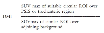
Indian Journal of Cancer, Vol. 47, No. 4, October-December, 2010, pp. 380-384 Symposium Exploring the role of FDG-PET in the assessment of bone marrow involvement in lymphoma patients as interpreted by qualitative and semiquantitative disease metabolic activity parameter PG Kand, BP Tiwari, S Basu, RV Asopa, UN Nayak Radiation Medicine Centre, Bhabha Atomic Research Centre, Tata Memorial Hospital Annexe, Jerbai Wadia Road, Parel, Mumbai 400 012, Maharashtra, India Code Number: cn10093 PMID: 21131749 DOI: 10.4103/0019-509X.73569 Abstract Bone marrow biopsy (BMB) is currently the standard method to evaluate marrow involvement in malignant lymphomas. However, there exist a number of pitfalls in this technique that can have important implications for initial staging, prognostification, and treatment of the disease. The present study was undertaken to investigate the utility of FDG-PET imaging in the detection of bone marrow involvement in untreated lymphoma patients. Forty untreated patients (36 males and 12 females) with either Hodgkin's disease (HD) (n = 17) or non-Hodgkin's lymphoma (NHL) (n = 31) underwent whole body FDG-PET study for disease evaluation. Bone marrow uptake of FDG was graded as absence or presence of disease activity at marrow sites by qualitative assessment. Semiquantitative analysis involved deriving disease metabolic index (DMI) using the following formula: DMI = SUV max of suitable circular ROI over PSIS or trochanteric region/ SUVmax of similar ROI over adjoining background. Findings of BMB and FDG-PET were compared for final analysis. Eleven out of 17 HD patients (12 males and 5 females) demonstrated concordance between FDG PET findings and BMB reports. Remaining 6 cases showed discordance of FDG-PET demonstrating presence of marrow involvement at marrow sites and uninvolved marrow on BMB. Twenty six of the 31 NHL cases (24 males and 7 females) demonstrated concordance between FDG PET findings and BMB reports. Remaining 5 cases showed discordance of FDG-PET demonstrating presence of marrow involvement at marrow sites and uninvolved marrow on BMB. All the BMB positive patients (2 of HD and 5 of NHL) demonstrated disease activity in bone marrow on FDG-PET study. All patients with absence of disease activity at marrow sites on FDG-PET scan (9 of HD and 21 of NHL) had histology proven uninvolved marrow. The quantitative assessment by DMI showed a mean of >2.5 in HD and NHL patients at the PSIS region and the trochanteric region bilaterally in cases of bone marrow involvement by the disease. FDG-PET is a useful adjuvant to BMB for the evaluation of bone marrow involvement in lymphoma patients. The disease metabolic index of >2.5 at the marrow sites can serve as a semiquantitative parameter for such diagnosis on FDG-PET in untreated patients of lymphoma.Keywords: Bone marrow biopsy, Hodgkin′s disease, FDG PET, Lymphoma, non-Hodgkin′s lymphoma Introduction Malignant lymphomas arise from neoplastic transformation of lymphoid cells in lymph nodes, spleen, or thymus gland. The disease is known to demonstrate a multifocal, hematogenous dissemination with bone marrow involvement. Being one of the curable forms of cancer, treatment and prognosis is significantly influenced by the histological subtype / grade, stage, and appropriate initial diagnosis. FDG-PET has been extensively evaluated for the staging, therapy monitoring, and surveillance of lymphoma patients. [1] Bone marrow involvement is considered an important factor in the staging and prognostication of malignant lymphomas, and hence, the examination of bone marrow (BM) is of great importance for the diagnosis and staging of the disease. [2] The incidence and clinical importance of marrow involvement varies according to various histological sub-types of lymphoma. The involvement of bone marrow in malignant lymphomas is seen commonly even at presentation in aggressive variants of non-Hodgkin′s lymphoma (NHL) while less commonly in Hodgkin′s disease (HD). Bone marrow biopsy (BMB) is the standard method to evaluate marrow involvement in malignant lymphomas. This standard diagnostic procedure, however, is associated with a high rate of false-negative findings, which may lead to inappropriate management.[3] Critical examination by generous BM biopsy samples can increase the diagnostic accuracy. [4] Also, the concentration of FDG in the bone marrow of lymphoma patients at initial pre-therapy imaging can reliably indicate lymphomatous involvement in the bone marrow, contributing to the assessment of the disease that presents a challenging task, and thereby initiation of correct treatment. The present study was undertaken to understand and investigate the utility of FDG-PET in the assessment of bone marrow involvement in untreated lymphoma patient. Materials and Methods Forty-eight patients diagnosed of malignant lymphoma referred to our Institute for whole body FDG-PET scan prior to treatment were included in the analysis. Whole body FDG-PET study was performed using standard imaging protocol. The tracer concentration within the bone marrow was assessed at the following sites for visual or qualitative evaluation: the spine, the pelvic bones (the bilateral iliac crest and region of posterior superior iliac spine), the proximal ends of the long bones (humerii and femora), and the ribs. The information from the scan was analyzed by qualitative and semiquantitative methods. Qualitative assessment The absence of tracer uptake or low-grade uptake (less than or equal to liver parenchyma intensity) in the marrow sites was interpreted as absence of disease activity. Tracer uptake of moderate to intense degree (more than liver parenchyma intensity) with associated heterogeneous or non-uniform (patchy) pattern was interpreted as presence of disease involvement [Figure - 1] and [Figure - 2]. Semiquantitative assessment This was performed by deriving Disease Metabolic Index (DMI) as a ratio of SUVmax (Gm-ml) values obtained from a suitable circular ROI over the posterior superior iliac spine (PSIS) - the usual site of BMB, the trochanteric region and the adjoining background (soft tissue in the gluteal and thigh region) in patients with bone marrow tracer uptake interpreted as presence of disease metabolic activity on qualitative assessment, i.e.,
BMB reports were obtained from the patient record sheets and compared with the FDG-PET findings [Figure - 3]. Results A total of 48 patients diagnosed of lymphoma (17 HD and 31 NHL) formed a part of the study. The mean age of the 17 HD patients (12 males and 5 females) was 29.82 ± 17.0 years. The mean age of the 31 NHL patients (24 males and 7 females) was 44.41 ± 18.0 years. BMB results from the case sheets of patients′ revealed involved marrow in 2 of the 17 HD patients and in 5 of the 31 NHL patients [Table - 1]. The qualitative assessment of the FDG-PET scan revealed findings suggesting presence of disease metabolic activity in 8 of the 17 HD patients and in 10 of the 31 NHL patients [Table - 2]. The comparison of BMB results with that of the FDG-PET results showed concordant findings in 64.7% of HD and 83.87% of NHL patients, whereas discordant findings in 35.29% of HD and 16.12% of NHL patients. All BMB-positive patients (2 of HD and 5 of NHL) demonstrated disease activity in bone marrow on FDG-PET study. All patients with absence of disease activity at marrow sites on FDG-PET scan (9 of HD and 21 of NHL) had histology proven uninvolved marrow. The quantitative assessment by DMI showed a mean of >2.5 in HD and NHL patients at the PSIS region and the trochanteric region bilaterally in cases with FDG-PET demonstrating disease activity at marrow sites and in cases of concordant and discordant findings [Table - 3] and [Table - 4]. In a single case from the concordant group significantly high values of DMI of 12 were noted, attributing to the extensive bone marrow disease. Discussion Evaluation of bone marrow disease is an important step in the work-up of patients with HD and NHL.[5],[6] The use of FDG-PET scan in the work-up of lymphoma patients has increased significantly with proven benefits in the staging, restaging, and prognostication of disease [7],[8],[9],[10],[11],[12] Despite the obvious role of FDG-PET in lymphoma there are contradictory reports on the accuracy of FDG-PET in marrow evaluation. [13] PET had an overall accuracy of 95% compared with 89% for bone marrow biopsy. [3] In another study of 42 patients, Jerusalem et al. demonstrated accuracy of detection for PET of only 39% (in biopsy confirmed cases) in indolent NHL. [14] A meta-analysis evaluating the ability of PET to identify bone marrow infiltration in (587 patients) found good (but not excellent) concordance particularly in HL and aggressive NHL. [15] In the routine examination of BMB there occur instances when it may not be possible to clarify the dilemma between a reactive lymphoid aggregate and neoplasia.[16] The determination of involvement of marrow may be difficult, given that no universally accepted standards exist. [17] This led us to explore more on the role of FDG-PET in the assessment of bone marrow evaluation in lymphoma patients. In our results, there was a 64.7% and 83.87% concordance noted in HD and NHL on comparison with the findings from BMB and FDG-PET. A higher discordance of 35.29% was noted in HD than 16.12% in NHL. In our observation, the FDG-PET scan had a high negative predictive value as all patients with absence of disease activity at marrow sites on the FDG-PET scan had histology proven uninvolved marrow. The pattern of the FDG uptake in the bone marrow sites is directly dependent on the pattern of infiltration of the marrow by the disease. Lymphomas may often manifest one or a mixture of the focal (involving one or more areas), diffuse (replacing the adipose tissue and hemopoietic elements), or dispersed (in the form of single or a limited number of neoplastic cells distributed in between the hemopoietic elements) patterns irrespective of the topographical location. [16] Bone Marrow hematoxylin-eosin (H and E) sections that appear to be uninvolved in the lymphomatous process may prove to be infiltrated by neoplastic cells being highlighted after immunohistochemistry. [18],[19] Therefore, we find that the challenges of reporting a BMB specimen extend to the interpretation of the FDG uptake in marrow sites in lymphoma patients. Pelosi et al. concluded from their study of 194 consecutive patients of malignant lymphoma that the diagnostic role of BMB and FDG-PET is complementary. [5] Pakos et al. in their meta-analysis of 587 patients have concluded that FDG-PET may complement BMB in the staging of primary and recurrent lymphoma. [15] Based of the criteria of FDG concentration seen in marrow sites with SUV >2.5, no reliable comparison with BMB could be sought by Elstrom et al. [20] Also, certain subtypes of indolent NHL are known to manifest lower FDG concentration. The semiquantitative analysis was performed taking into account the complementary nature of both the investigations, mixed reports on the reliability to accurately detect infiltration of marrow on FDG-PET, and the challenges of both the investigations. In the group of patients with positive BMB and FDG demonstrating disease activity in bone marrow sites, our results revealed a mean DMI of > 2.5 at multiple sites. Hence, a lower threshold of 2.5 for DMI can be more suitable to guide the biopsy sites in FDG concentrating bone marrow disease infiltration. We believe that DMI may be utilized as a reliable marker for the disease activity in difficult situations as it takes into account the incremental metabolism and GLUT receptor overexpression above the basal metabolism and basal GLUT receptor expression of the tissue of interest and its adjoining background in the same patient. This can help targeting of BMB to identify the areas of sampling within a site, and thereby, improving the quality of the sampling. FDG-PET imaging findings can thus be exploited for the diagnosis of bone marrow involvement in lymphoma, particularly if the involvement is distant from the usual biopsy site and to guide the generous collection of samples from the highest metabolically active areas that should ideally be the intended biopsy site. However, these findings may not be applicable in patients post chemotherapy or post administration of granulocyte colony-stimulating factors (GM-CSF) due to their effect on increasing FDG uptake in the marrow due to marrow stimulation. Conclusion FDG-PET can be a very useful complimentary technique to the standard BMB for evaluating bone marrow involvement in lymphoma patients. Both qualitative and semiquantitative DMI can be utilized for targeting a judicious BMB. References
Copyright 2010 - Indian Journal of Cancer The following images related to this document are available:Photo images[cn10093t2.jpg] [cn10093t3.jpg] [cn10093t1.jpg] [cn10093f2.jpg] [cn10093f1.jpg] [cn10093f3.jpg] [cn10093t4.jpg] |
| |||||||||


![[Figure - 1]](/showimage?cn/photo/cn10093f1.jpg)
![[Figure - 2]](/showimage?cn/photo/cn10093f2.jpg)
![[Figure - 3]](/showimage?cn/photo/cn10093f3.jpg)
![[Table - 1]](/showimage?cn/photo/cn10093t1.jpg)
![[Table - 2]](/showimage?cn/photo/cn10093t2.jpg)
![[Table - 3]](/showimage?cn/photo/cn10093t3.jpg)
![[Table - 4]](/showimage?cn/photo/cn10093t4.jpg)