 |
| About Bioline | All Journals | Testimonials | Membership | News |
|
||||||
|
||||||
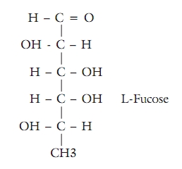
Indian Journal of Cancer, Vol. 47, No. 4, October-December, 2010, pp. 452-457 Review Article Serum fucose level in malignant diseases NG Sawke, GK Sawke Department of Pathology, People's College of Medical Sciences and Research Center, Bhopal, M.P., India Correspondence Address: Code Number: cn10105 PMID: 21131761 DOI: 10.4103/0019-509X.73549 Abstract We review the present knowledge of serum fucose with special attention to its relation with various malignant diseases. We summarize the role of serum fucose as a useful diagnostic and prognostic marker when used singly or in combination. The purpose of this review is to provide an expert opinion on the practical and applied aspect of serum fucose level in clinical practice and research settings. Our review is based on information from published research studies, library books, and electronic searches through Medline and PubMed. The available published data were used as the basis for recommendations. Each of the subsections concludes to provide information to assist the clinicians and the research scientists make informed decisions.Keywords: Malignant, prognostic, serum fucose level Introduction The glycoproteins are widely distributed in animal tissue. Fucose is the only sugar which is present in L-form. All the other sugars are present in the D-form, and include D-glucose, D-galactose, D-manonose, D-glucosamine, and so on. Glycoproteins are compounds composed of proteins and carbohydrates in which the sugars are firmly linked to the peptide portion of the protein. Elevated serum levels of protein-bound fucose have been reported in patients with malignant tumors. The present study is undertaken to find out the diagnostic significance of serum fucose in various malignant tumors as diagnostic and prognostic markers. For a long time, it has been known that methylpentoses are the constituents of polysaccharides of plant and bacterial origin. Fucose was first found in animal bodies as a constituent of blood group substances, combined with other sugars. The structural formula of L-fucose is:
Glycoproteins comprise of a large number of substances of diverse biological importance and behavior, namely: antibodies, enzymes, hormones, blood group substances, mucus secretions, membranes, and collagens. The levels of glycoproteins are elevated in a number of diseases including cancer. [1] Their place in health and disease has been reviewed periodically. Elevated serum levels of protein-bound fucose have been reported in patients with malignant tumors by Macbeth and McBride,1965; Rosato, 1968; 1971. [2],[3],[4] Reports about serum fucose in malignancy have appeared in our country also, with special reference to various malignancies. The present study is undertaken to find out the diagnostic significance of serum fucose in various malignant tumors. Biochemistry of Glycoproteins Glycoproteins as a group, have multiple and complex functions. The early work in this field was done by Grevenstuk 1929. and also by Remington 1933. They studied the chemistry of these components and their physiological significance. According to the recommendations of the American Physiological Society and The American Society of Biochemists, the compound of a protein molecule, with a substance or substances containing a carbohydrate group, other than nucleic acid, was classified as a glycoprotein. Meyer has suggested that glycoproteins are of two general types, one having the proteins bound to the carbohydrate through polar linkages, which can easily be split by a concentrated salt or alkaline solution, known as ′mucoproteins′, and another one with firmer, presumably covalent bonds, between the protein and carbohydrate. Little is known about the site of formation of the serum glycoproteins. However, various views have been proposed by several workers in support of an increase in serum glycoproteins in malignant and other diseases. It is suggested by Seibert FB, Seibert MV, A. Atno AJ, and Campbell HW that elevation of glycoproteins above the normal levels reflects processes of tissue destruction at the site, and release of preformed glycoproteins from the tissue, or it may be due to local synthesis and liberation of glycoproteins by tumor cells. [1] Shetlar MR, Foster JV, and Kelly KH are of the view that the increased serum glycoproteins level in diseases reflects processes associated with tissue proliferation rather than with tissue destruction. [5] Fucose The presence of fucose in serum glycoproteins has been demonstrated by Waldron and Woodhouse. Dische et al. also established that glycoproteins contain galactose, mannose, glucosamine, galactosamine, sialic acid, and so on, other than fucose. [6] Fucose is the only sugar that is present in the L-form. L-fucose behaves differently in the photoelectric field and so it can be estimated easily by using colorimetric methods. A specific color reaction of methylpentoses, a spectrophotometric micromethod for their determination, was first described by Dische Z, and Shettles LB. [7] Determination of L-Fucose Serum protein-bound fucose was estimated by the method of Dische and Shettles, utilizing the modification proposed by Winzler. L-Fucose was estimated according to the method of Winzler, using cysteine hydrochloride. L-Fucose was assayed by dissolving ethanol-precipitated proteins of serum in alkali, heating with sulfuric acid, and determining the color after addition of cysteine. Standard L-Fucose was procured from the Sigma Chemical Company, MO, US. The color produced by hexoses under these conditions was corrected by determining the absorbance at 400 nm and 430 nm. [8] At present, the Biotechnology Research Laboratories have devised a kit to use, with automated analyzers, for the assay of L-fucose. T Sakai, K Yamamoto, H Yokota, K Hakozaki-Usui, F Hino, and I Kato, developed this kit for a simple enzymatic assay of free L-fucose in serum and urine, by means of a newly isolated L-fucose dehydrogenase, which could be used with automated analyzers. They measured L-fucose in healthy subjects, cancer patients, and patients with other diseases. The automated analyzer was linear up to at least 3.0 mmol of L-fucose per liter. To evaluate the sensitivity, they assayed an L-fucose sample 20 times. The standard deviation was < 7 micro mol /L. This assay measured the L-fucose specifically. The Results were not affected by D-fucose, D-galactose, L-arabinose, D-glucose, DL-mannose, L-lyxose, D-xylose, L-rhamnose, L-sorbose, D-fructose, N-acetyl-D-glucosamine, N-acetyl-D-galactosamine, N-glycolyl neuraminic acid, or N-acetyl neuraminic acid, even in a concentration of 10 mmol /L. Analytical recovery of added L-fucose was good (90 - 100%), even at low concentrations (25 micro mol /L). [9] Normal Fucose Levels Serum protein-bound fucose levels have been estimated by different workers in healthy control subjects. Winzler RJ in his study has noted the normal serum fucose level in 20 healthy controls. It was found to be 8.9 ± 0.6 mg / 100 ml. [9] Macbeth RAL and McBride G. reported the normal fucose level to be 8.2 ± 0.6 mg / 100 ml and Macbeth and McBride, in 1965, reported the normal fucose levels to be between 7.5 mg and 8.4 mg%. [2],[10] Rosato et al. observed that normal serum fucose level was below 12 mg% in healthy controls. However, in their second study they found the normal value of serum fucose to be less than 3.35 X 10 -3 mg fucose / mg protein.[3] Sharma and Sur tested the serum fucose sialic acid levels in Indian children and adults. They found the normal serum fucose levels to be 3.87 ± 0.08 mg% in children and 4.6 ± 0.13 mg% in adults. [11] Spiro RG in his study found the normal serum fucose level to be 8.6 mg%. [12] Deyasi, Aikat and Sengupta found the normal serum fucose level to be between 6 mg and 13 mg% from the sera of 50 normal individuals, who served as the control. [13] Mehta and Venkatraman showed the normal serum fucose level to be between 6.04 and 15.51 mg%. [14] Pradhan et al. reported the normal serum fucose levels in 50 normal healthy individuals. It was found to be 10.5 mg% (range: 3.5 to 17 mg%). [15] Kaswan HS and Kaushik SK studied 20 healthy individuals for normal serum fucose level. In the 20 healthy controls, the serum fucose level ranged between 3.42 and 9.05 mg% with an average of 6.05 ± 2.99 mg%. [16] Lodha et al. conducted a study on 25 healthy control subjects for normal serum fucose level. The normal serum fucose level was found to be between 4.87 mg and 16.18 mg% with an average of 8.57 ± 2.64 mg%. [17] Kadu AR and Deshraj BP, in their study took 20 normal healthy controls. They found that the normal serum fucose level ranged between 3.28 and 9.24 mg% in their study of normal healthy controls. [18] Serum Fucose Level in Malignancies In 1955, Winzler and colleagues reported a significant increase in serum protein-bound fucose in patients with advanced cancer and other diseases. In their study of 15 cases of advanced cancer they found the serum fucose levels to be 14.2 ± 2.1 mg% in comparison to the normal level of 8.9 ± 0.6 mg%. [9] Rosato, in his study, noted that when the serum fucose levels were correlated with total serum proteins, the patients with malignant disease had a serum fucose level above the significant dividing point, 3.35 X 10 -3 mg fucose / mg protein; while, 70% of the benign conditions had a value below 3.35 X 10 -3 mg fucose / mg protein. Rosato studied 150 patients in the series, 46 proved to have breast carcinoma, 70% of the patients with carcinoma had elevated serum fucose level, whereas, only 18% of the patients without malignancy had elevated levels. The serum level of 3.35 X 10 -3 mg fucose / mg protein was taken as the dividing point. [8],[9] A total of 51 patients had elevated serum fucose levels, and of these, 63% had carcinoma of the breast. They further considered these 51 patients with respect to age, an increase was seen in the correlation between positive results (i.e., 3.35 X 10 -3 mg fucose / mg protein) and the eventual findings of cancer. These data were re-examined by using new criteria, wherein a patient was diagnosed as having carcinoma serologically, if the serum fucose was 25% or higher and serum protein was less than 8.5 gm%. Using these criteria they found that 67% of the patients with carcinoma had elevated levels, whereas, only 13% of the patients with benign disease had elevated levels. A total of 44 patients had satisfied these criteria and 70% of these would subsequently be shown to have breast carcinoma as compared to 63%, when the old criteria were used. They again considered the group of patients in whom the tests had given positive results, by age group, and got an even better marked correlation between the older age groups and the percentage of positive results. [8],[9] However, they proposed that the evaluation of the data by age groups was of greater significance. With both the older criteria and the new criteria, the significance of positive results became more meaningful as the age considered increases.This was due to the fact that many of the false positive results were obtained in younger population groups, in whom the incidence of carcinoma was low. [3],[4] Arya DB and Bhatnagar KK estimated the serum fucose levels in breast lesions. This study was undertaken in 51 cases with breast lumps. All the patients were subjected to a detailed clinical examination. The cases for study were divided into two groups; Group - I: Patients with breast lump; Group - II: 20 control subjects. The serum fucose levels in the 20 control cases (both males and females) ranged from 4.0 to 7.2 mg% and the average serum fucose level was 4.92 ± 0.87 mg%. In 10 cases with benign breast lesions, it was 6.15 ± 1.78 mg%, and in the 41 cases with malignant lumps the average level was 8.09 ± 1.44 mg%. The difference between control and benign, control and malignant, and benign and malignant, were subjected to the ′t′ test for statistical significance, and it was found that the rise in the serum fucose level from normal to benign cases was ′possibly significant′, while the rise in the level from the benign to malignant group was ′highly significant′. The rise in the level from the normal to the malignant group was also ′highly significant′. Out of 41 cases with malignant lumps, 37 patients (90.2%) had a serum fucose level of above 6.15 mg%, that is, above the average levels of benign cases and controls. They also mentioned that there was a constant rise of serum fucose levels with the progression of stages in breast cancer (Manchester Classification). They found that the rise in serum fucose level in malignant cases was directly proportional to the stage of breast cancer and inversely proportional to the ages of the patients. The serum fucose level tended to decline slightly after the removal of the tumor mass, more in cases without metastasis than in those with metastasis. However, the decrease in level was not significant. [19] Deyasi SK, Aikat BK, and Senguta U, have estimated serum fucose in various malignant lesions. They have studied 250 sera of cancer patients. Eighty percent of the sera showed elevated serum fucose levels (above 13 mg / 100 ml) compared to a range of 6 to 13 mg / 100 ml found in the normal sera. [13] [Table - 1] To show the correlation among the carcinoembryonic antigen (CEA), Reagin isoenzyme, and serum fucose level, Deyasi et al. studied the sera of 250 cancer patients. In this study they found a positive correlation between the raised fucose levels and CEA positivity. All the CEA-positive sera showed a fucose level above 13 mg%. The positivity of the Regan isoenzyme in these 250 sera, was 16.4%, irrespective of the organ involved.[20] Dutta TK, Sengupta U, and Gupta BD studied the serum protein-bound fucose in malignant tumors. They observed a uniform elevation of serum fucose levels in a group of patients with common malignant tumors. In their study, they found that the mean serum fucose level was the highest in the carcinoma of the cervix and next in order were those in carcinoma breast, lymphoma, and oral cancer. After treatment by radiotherapy, the level of serum fucose did not always come down to the normal range. [21] Solanki RL, Ramdeo IN, and Sachdeo KN estimated the protein-bound fucose levels with healthy control subjects and benign and malignant lesions of the breast. The mean value of the control group was 6.5 mg ± 1.46 mg%. In benign lesions of the breast, the mean value was 11.55 mg ± 1.15 mg%. In malignant lesions of the breast, the mean value was 19.58 mg ± 0.86 mg%. The serum fucose showed a constant rise with progression of the disease. [22] Pradhan et al. analyzed a total of 220 sera, obtained from patients suffering from various malignant diseases, for serum fucose levels. The normal serum fucose levels in 50 normal individuals were found to be 10.5 mg% (range: 35 to 17 mg%), and 172 cases of cancer had elevated fucose levels, ranging from 14 to 40 mg% with an average of 19.86 mg%. The lowest values (15.2 mg%) were seen in malignant lesions of the mastoid region and the highest (32.5 mg%) in cancer of the pancreas, keeping 19.86 mg% as the positive percentage value. They suggested serum fucose level as a useful adjunct in the diagnosis of various malignant diseases. [15] [Table - 2] Kaswan HS and Kaushik SK, studied 70 patients with lump in breast and 20 healthy individuals for serum fucose levels. The SFL ranged between 3.42 and 9.05 mg% with an average of 6.05 ± 2.99 mg%. Eighteen benign cases had preoperative fucose levels ranging between 8.06 and 13.5 mg% with an average of 10.78 ± 2.42 mg%. These benign cases, on the tenth postoperative day, showed fucose levels ranging between 7.78 to 13.20 mg% (average 10.37 ± 1.64 mg%). There was no significant difference between the pre- and postoperative fucose levels, but both these levels differed significantly from the normal fucose levels in the control group. In the 52 cases with malignancy of the breast, the pre-operative fucose levels ranged between 12.99 and 23.92 mg% with an average of 18.72 ± 5.598 mg%. On the tenth postoperative day, the levels ranged between 12.22 and 21.46 mg% with an average of 17.03 ± 5.522 mg%, suggesting that there was no significant difference in the pre - and postoperative levels. They also tried to establish the relationship between serum fucose level and age of patients, but could not get any positive correlation. [16] Lodha SK, Kalra VB, Sarin PN, Utreja SR, and Mathur JS, completed a study of 100 sera, from patients admitted with malignant tumor and 25 healthy controls, for the serum fucose levels. The serum fucose levels were markedly raised in cancer subjects and were statistically very significant (P<0.001) when compared with those of the control subjects. They reported that the mean serum fucose in 68 cases of cancer, with definite evidence of metastasis, was significantly higher than in the cancer subjects without established metastasis. [17] Mishra VK, Basu PK, Saxena SP, and Om Prakash showed the significance of serum fucose levels in conglomeration with Trans-sternal phlebography in the management of breast cancer. The study was conducted on 63 subjects with breast diseases. Serum fucose levels of these subjects and 22 normal healthy subjects were determined. The serum fucose levels increased in both benign and malignant breast disease; the magnitude being more in the latter. The serum fucose level increased with the stage of malignancy and decreased postoperatively at all stages of cancer of breast, as well as in benign breast disease.[23] Kadu AR and Deshraj BP estimated serum fucose levels in 130 patients with different breast lesions (50 benign and 80 malignant) and 20 normal controls, to assess its value, in judging the response to treatment and for detection of recurrences; and patients resistant to chemotherapy / surgery. The serum fucose levels ranged between 3.28 and 9.24 mg% (mean 5.93 ± 1.73 mg%) in healthy individuals as compared to 5.12 and 11.26 mg% (mean 7.78 ± 1.78) in patients with benign lesions, and 12 mg and 28.50 mg% in patients with malignant lesions (mean 18.08 ± 4.89 mg%). The value in the malignant group was significantly high when compared with those in the control and benign groups. No. correlation was found between the age and serum fucose level in the healthy as well as the benign and malignant groups of patients. Approximately 65% of the patients with breast carcinoma had serum fucose levels exceeding 3.35 x 10 -3 mg fucose / mg protein, while all the patients with benign lesions showed values below this level. The rise in serum fucose level (SFL) was observed with the progression of malignancy from stage I to IV and mean SFL was significantly higher in patients with metastasis. Seventy percent of the patients who responded to treatment showed a fall in SFL in the post-treatment period. Furthermore, all patients who developed recurrences, local or distant, showed persistently high SFL in the post-treatment period. [18] Fernandez-Rodriguez J; et al. determined fucose level in the sera and in tumors of colorectal adenocarcinoma patients. Total, free, and bound fucose were significantly higher (P < 0.001) in tumoral tissue. When total and bound fucose were normalized to total protein (nmol / mg protein), an elevation in both parameters was found in colorectal cancer patients, although only that for total fucose was statistically significant (P < 0.001). [24] Conclusion Serum fucose level (SFL) has been estimated by different workers in healthy and cancer subjects. Most of them have found significantly elevated SFL in subjects with malignant diseases as compared to healthy controls. A marked correlation between the older age group of cancer patients and raised SFL was observed, with the conclusion that SFL could be a good screening test singly or in combination with other relevant markers in subjects suspected to have cancer, with advanced age. It is also observed that the mean SFL obtained in cancer patients with metastasis and in advancing stages were significantly higher than the subjects without metastasis, hence it can be used as a useful prognostic indicator. A large study should be undertaken in a prospective manner, to further strengthen and validate the data. References
Copyright 2010 - Indian Journal of Cancer The following images related to this document are available:Photo images[cn10105t1.jpg] [cn10105t2.jpg] |
| |||||||||

![[Table - 1]](/showimage?cn/photo/cn10105t1.jpg)
![[Table - 2]](/showimage?cn/photo/cn10105t2.jpg)