 |
| About Bioline | All Journals | Testimonials | Membership | News |
|
||||||
|
||||||
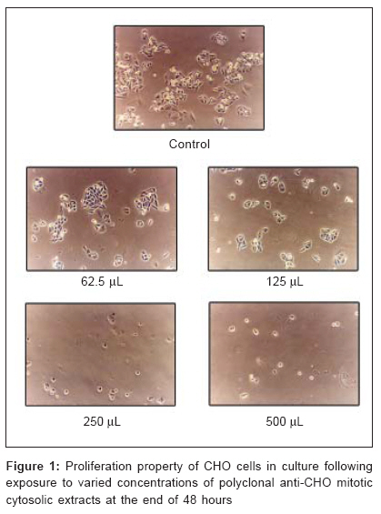
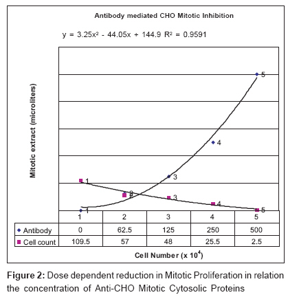
Journal of Cancer Research and Therapeutics, Vol. 2, No. 3, July-September, 2006, pp. 126-128 Original Article Polyclonal antibody-mediated mitotic inhibition in Chinese hamster ovary cells Maddaly R, Deepa PV, Pai GM, Preetha B, Ghosh S, Paul SFD Department of Human Genetics, Sri Ramachandra Medical College and Research Institute (Deemed University), Porur, Chennai Code Number: cr06028 Abstract The nucleus of a mammalian cell undergoes profound reorganization when the cell enters mitosis and a number of proteins involved at various levels of the cell cycle have been characterized. The presence of mitotic-specific proteins has been reported and their roles are important in understanding the mechanics of cell division. The ability of antibodies to recognize mitotic protein antigens and further inhibit mitosis is potentially valuable in their role as therapeutic and diagnostic agents in cancer therapy. In this study, we have aimed to analyze proteins isolated from mitotic cells of Chinese hamster ovary (CHO) cells and their significant role in inhibiting mitosis. The proteins extracted from mitotic cells were processed and antibodies produced. It was observed that the secondary response that yielded an antiserum of 1:8 titer was predominantly IgG. The antiserum was effective in inhibiting mitosis in CHO cells in culture in a dose-dependent manner. Although inhibition of mitosis was apparent by cell proliferation studies, there was no apparent effect of the antiserum on other cell morphology and culture characteristics. The unique molecular structure of the antibody by which it bivalently binds to a broad array of antigenic epitopes serves as the foundation of its utility. These antibodies, being polyclonal in nature, are targeted against a whole range of proteins; and their multiple epitopes involved in process of cell division might hence mediate recognition or inhibition of function of such proteins in a wholesome manner and thus accomplish inhibition of mitotic progression.Keywords: CHO mitotic cells, extracts, mitotic inhibition, polyclonal antibodies Introduction The nucleus of a mammalian cell undergoes profound reorganization when the cell enters mitosis. The nuclear envelope breaks down and the chromatin condenses into discrete chromosomes. A number of proteins involved at various levels of the cell cycle have been characterized and a variety of protein kinases have been separated and identified in extracts from mitotic and interphase cells.[1] Certain nonhistone proteins, possibly phosphoproteins, of mitotic cells are not present in interphase cells. The presence of mitotic-specific proteins has been reported by using two-dimensional gel electrophoresis.[2] Protein profiles in the various phases of the cell cycle revealed that in normal cell cycle, an acidic protein of 37 KDa was specifically associated with chromosome condensation at the mitotic phase.[3] It was also found that the condensation factor was cytoplasmic, which enters the nuclear membrane causing condensation of chromatin during mitosis.[4] Protein profile during cell cycle is critical for the various cellular mechanisms and proper regulation of the same. The roles of proteins unique to the mitotic cells are important in understanding the mechanics of cell division. Antibodies are proving to be very useful as diagnostic, imaging and therapeutic reagents in clinical medicine. Recognition of such proteins by antibodies and inhibition of their function (during mitosis) is extremely important in the study of cell cycle kinetics. The ability of such antibodies to inhibit mitosis is potentially valuable in their role as therapeutic and diagnostic agents in cancer therapy. The role of a given protein during mitosis can be determined effectively by two methods. One powerful approach involves induction of mutations that alter gene(s) encoding a protein of interest. Though this method allows one to examine the function of a protein with precision, the requisite mutant cells must be found; and the list of mitosis-blocking mutations is not very extensive at present. An alternative approach involves the microinjection of antibodies against putative mitotic proteins into dividing cells. The microinjection method is advantageous because the antigen(s) recognized by the antibody need not be well characterized at the initiation of the experiments. Also, the effects of the antibodies can be monitored in real time.[5] In this study, we have aimed to analyze proteins isolated from mitotic cells of Chinese hamster ovary (CHO) cells and their significant role in inhibiting mitosis by treating CHO cells in culture with polyclonal antibodies raised against purified whole-cell mitotic extract and also analyze the recognition of proteins unique to mitotic cells by antibodies thereby effectively inhibiting progression to mitosis in such cells. Materials and methods CHO cell lines were cultured in Dulbecco's Modified Eagle's Medium (GIBCO) containing 15% FBS at 37°C in humidified atmosphere containing 5% CO 2 . Cells were synchronized at mitotic phase by addition of 75 µL of colcemid (GIBCO) and incubated for 2 h. A maximum number of mitotic cells were harvested by selective detachment. Mitotic cytosolic proteins were extracted using ReadyPrepT Sequential Extraction Kit (Bio Rad) containing Tris base. The mitotic cell pellets obtained by selective detachment and washing with medium twice were treated individually with 1 ml cold ReadyPrep reagent 1 (which extracts only the most soluble proteins, such as cytosolic proteins) and the pellet was mechanically agitated using Teflon-coated pestle and centrifuged at 1,000 rpm; 10 min. The supernatant containing the proteins was aspirated and a buffer exchange was performed by dialysis with 1X PBS at 4°C overnight. The proteins extracted from mitotic cells were processed and purified by sephadex gel filtration using 1X PBS as the wash buffer. The concentration of the proteins in the whole-cell mitotic extract following purification by gel filtration was estimated using Bradford protein assay. One healthy previously unimmunized rabbit was given a subcutaneous injection of 1 ml sterile soluble mitotic extract (filtered through 0.22µ filter) emulsified with an equal volume of complete freund's adjuvant at multiple sites. The rabbit was given two booster injections of 1 ml (emulsified with incomplete freund's adjuvant) subcutaneously on 17th and 25th day after first immunization. Blood samples were collected on the 7th, 24th and 32nd day after the first immunization, allowed to clot at room temperature and antiserum collected. The antiserum thus collected was tested by countercurrent electrophoresis to confirm the presence of antibodies for CHO mitotic extract. The titer of the antiserum was established by conventional Agarose double diffusion. Purification of IgG from whole serum was done by Proteinase A column with phosphate buffered saline as the buffer. Thus, three sets of reactants - whole serum, serum devoid of IgG and purified IgG - were obtained and were individually tested for reactivity with CHO mitotic extracts. Mitotic inhibition assay with CHO cells Interphase CHO cells were plated at a seeding density of 11,700 cells per culture plate. Five such plates were seeded, with one of them as control and the other four supplemented with the polyclonal anti-mitotic protein whole serum of four serial dilutions from 500 to 62.5 µl. Test control plate with the same cell seeding density with 500 µl of serum from unimmunized healthy rabbit was included in the study. The plates were incubated for 48 h, trypsinized and cell counts taken from individual culture plates. The mitotic inhibition property of the polyclonal antibody was assessed by constructing a cell proliferation index after an incubation period of 48 h in the presence of varied concentrations of the same. The morphology and other properties of the cells in culture were observed and photo-documented. Results Protein quantification of CHO mitotic extracts showed values of 0.6 mg/ml. Serum isolated following immunization showed presence of antibodies for whole-cell CHO mitotic extract and its titer was established as 1:8 after the second booster. The IgG fraction isolated by affinity chromatography and tested by immunodiffusion for its contribution to the reactivity of the antiserum showed distinct precipitin line after concentration by solid sucrose dialysis on comparison with the same before concentration (in which reaction was absent). The serum devoid of IgG did not show reactivity, thus indicating a secondary antibody response containing predominantly IgG. Forty-eight hours further to incubation in the presence of the polyclonal anti CHO-mitotic extract antibodies, the plates were trypsinized and total cell counts measured. The control showed a cell count of 109.5 x 10 4 and plates incorporated with 62.5, 125, 250 and 500 µL of the antiserum showed total cell counts of 57 x 10 4, 48 x 10 4, 25.5 x 10 4 and 2.5 x 10 4 respectively [Figure - 1]. Although there was a dose-dependent reduction of cell numbers further to the incubation [Figure - 2] there were no significant changes in the morphology of either the cell characteristics or culture characteristics such as medium quality. The test control did not show any significant difference from that of the control. Discussion A number of proteins involved at various levels of the cell cycle have been characterized and a variety of protein kinases have been separated and identified in extracts from mitotic and interphase cells.[1] The presence of mitotic-specific proteins has been reported by using two-dimensional gel electrophoresis.[2] Protein profile during cell cycle is critical for the various cellular mechanisms and proper regulation of the same. The roles of proteins unique to the mitotic cells are important in understanding the mechanics of cell division. Antibodies have provided and will continue to provide scientists and clinicians an extraordinarily powerful tool for use in the research laboratory and clinic. The unique molecular structure of the antibody by which it bivalently binds to a broad array of antigenic epitopes serves as the foundation of its utility. Antibodies are 'tag' molecules commonly used, both in vitro and in vivo, for a diverse array of functions that include identification, isolation, modification and destruction. Antibodies can directly mediate effector functions such as cytotoxicity or can be used to deliver effectors such as radionuclides, immunotoxins or enzymes, which can be bound to the molecule. Inhibition of mitotic progression by microinjection of monoclonal antibody to mitotic microtubule-associated protein isolated from CHO cells either as ascites fluid or IgM has been demonstrated.[5] In the present study, polyclonal antibodies were raised against an entire range of cytosolic mitotic proteins with an aim to demonstrate the ability of such an antiserum to inhibit mitosis. However, experiments include subjecting CHO cell cultures with sera from unimmunized rabbits, which did not show much deviation from normal cultures but showed a marked difference from cultures exposed to antiserum containing polyclonal antibodies to whole mitotic cellular cytoplasmic extracts. The fact that cells in interphase and mitotic phases can have common proteins predominantly, and the aim being to obtain a 'net-inhibitory' effect through multiple epitopes; has led us to concentrate on polyclonal antibodies to whole mitotic cytosolic protein extracts and the applicability of the same in inhibiting mitotic progression. Further, the rationale for usage of sera from healthy, previously unimmunized rabbits as test controls, is that the difference in control and antiserum lies only in the antibodies of the IgG class being present in the antiserum; all other properties of the test control and test serum being utmost similar. Although cell division was inhibited, no further damages to the cells were observed; and the cells in presence of the varied concentrations of antibodies exhibited normal healthy morphology. Although the structure, functions and properties of antibodies are well understood along with the essential principles of Antigen-Antibody interactions, our study demonstrates inhibition of progression of mitosis in cultured CHO cells. Inhibition of mitosis mediated by polyclonal antibodies is not only an important tool in studying cell cycle kinetics but also has potential applications in the field of cancer immunotherapy. These antibodies, being polyclonal in nature, are targeted against a whole range of proteins and their multiple epitopes involved in process of cell division and hence mediate recognition or inhibition of function of such proteins in a wholesome manner and thus accomplish inhibition of mitotic progression. Also the contribution of the IgG class of immunoglobulins in the antiserum to its reactivity with the antigen (CHO mitotic extract) has been demonstrated in the present study. The uses of polyclonal antibodies, which are relatively easy to produce in a timely and cost-efficient way compared to monoclonal antibodies, prove to be advantageous in such a scenario. References
Copyright 2006 - Journal of Cancer Research and Therapeutics The following images related to this document are available:Photo images[cr06028f1.jpg] [cr06028f2.jpg] |
| |||||||||
