 |
| About Bioline | All Journals | Testimonials | Membership | News |
|
||||||
|
||||||
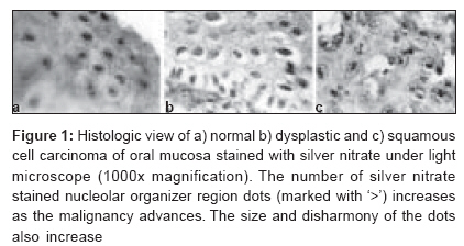
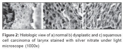
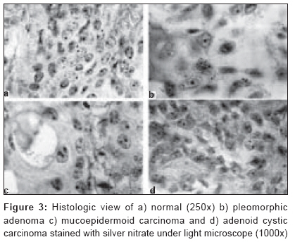
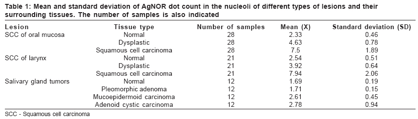
Journal of Cancer Research and Therapeutics, Vol. 2, No. 3, July-September, 2006, pp. 129-131 Original Article Diagnostic value of silver nitrate staining for nucleolar organizer regions in selected head and neck tumors Eslami Behnam, Rahimi Hessam, Rahimi Farzaneh, Khiavi MonirMoradzadeh, Ebadifar Asghar Basic Sciences Research Dept, Iran Center for Dental Research, Tehran Code Number: cr06029 Abstract Background: The present study is aimed to assess the usefulness of silver nitrate staining of nucleolar organizer regions (NORs) as a quantitative criterion for the diagnosis of selected head and neck tumors.Materials and Methods: The silver nitrate staining technique was used on 195 paraffin blocks collected from 85 patients. The samples consisted of 21 squamous cell carcinoma (SCC) of larynx, 28 SCC of oral mucosa and 36 samples of most common salivary gland tumors. Mann-Whitney U-Test was used for data analysis. Results: A significant difference was seen in the number of AgNOR dots between oral and laryngeal SCC with surrounding dysplastic and normal tissues ( P < 0.001) and also between mucoepidermoid carcinoma and adenoid cystic carcinoma with pleomorphic adenoma and normal salivary gland tissue ( P < 0.001). Conclusion: The silver nitrate staining for NORs is a useful method for aiding the diagnosis of malignant and dysplastic mucosal lesions and also malignant and benign salivary gland tumors. Keywords: Laryngeal squamous cell carcinoma, malignancy, nucleolar organizer regions, oral squamous cell carcinoma, salivary gland tumors, silver nitrate staining Introduction Nucleolar organizer regions (NORs) are loops of DNA that encode ribosomal RNA and are considered important in the synthesis of proteins. NORs can be selectively stained by a silver colloid technique and can be visualized as black dots under the transmission microscope. These visualized structures are commonly termed AgNOR dots.[1] The number of AgNORs rises with the increasing proliferative activity of cells.[2] Thus, compared to normal or benign lesions, the number of AgNOR dots in malignant lesions is higher.[3] Due to its simple, quick and convenient nature, this method has been used in histopathologic diagnosis of various benign and malignant human tumors, such as breast, prostate and salivary gland tumors.[4],[5],[6],[7] In this study, we evaluated the diagnostic value of silver nitrate staining of NORs in SCC of larynx and oral mucosa and also common oral salivary gland tumors. Materials and methods In this descriptive cross-sectional study, we used the silver nitrate staining technique[8] on 195 paraffin blocks collected from 85 patients from the archives of Taleghani Hospital (Tehran, Iran). The silver nitrate solution used for staining the samples was produced by mixing 2 g of gelatin in 100 ml of 1% formic acid with two parts of 50% silver nitrate solution in distilled water. The samples consisted of 21 SCC of larynx and 28 SCC of oral mucosa and their surrounding normal and dysplastic tissues, including 36 samples of the most common salivary gland tumors consisting of 12 pleomorphic adenomas, 12 mucoepidermoid carcinomas and 12 adenoid cystic carcinomas with their surrounding normal salivary gland tissues. AgNOR dots were counted by a pathologist with the aid of a graticule using conventional light microscopy with 1000x magnification, assessing the nuclei of 100 cells randomly selected from 10 distinct regions per slide [Figure - 1][Figure - 2][Figure - 3]. Then the nonparametric Mann-Whitney U-test was performed to analyze the data. Results The results are shown in [Table - 1]. The statistical analyses performed with Mann-Whitney U-test showed a significant difference in the number of AgNOR dots between oral and laryngeal SCC with dysplastic and normal surrounding tissuesin oral and laryngeal SCC when compared with dysplastic and normal surrounding tissue ( P < 0.001). The dysplastic surrounding tissue also had a significant difference in the number of AgNOR dots when compared with normal tissue ( P < 0.001). In both types of lesions, the number of AgNOR dots in cancerous area was almost two times higher than the normal tissue and 1.5 times higher than surrounding dysplastic tissue. A significant difference in the number of AgNOR dots was also seen between mucoepidermoid carcinoma and adenoid cystic carcinoma with pleomorphic adenoma and normal salivary gland tissue ( P < 0.001). There was also a significant difference between the number of AgNOR dots in the malignant salivary gland tumors and mucosal squamous cell carcinoma ( P < 0.001). The results also showed that the AgNOR dots in normal tissue were rounded and small and usually couldn't be detected outside the nuclei, while in the cancerous lesions, the shape of the dots varied and their harmony and equivalency was lost. In malignant lesions, the dots also seemed to localize in one location with increased size and color [Figure - 1][Figure - 2][Figure - 3]. Discussion Qualitative and quantitative changes of AgNORs may provide useful information about nucleolar activity in hyperplastic and neoplastic conditions.[9] Although various techniques for evaluating AgNORs have been employed, including enumeration, measurement of surface area and analysis of their distribution patterns, the counting of AgNOR dots is the most widely used method due to its simplicity and easy reproducibility.[10] Variations in size and/or number of AgNOR dots might depend on the stage of the cell cycle, transcriptional and metabolic activity of the cell or the number of NOR-bearing chromosomes in the kariotype.[10],[11],[12] In a rapid proliferative cell, the chromosomal and AgNOR distribution remains disorganized with the resultant formation of multiple, small and dispersed nucleoli. Actively proliferating cells have impaired nucleolar association and therefore exhibit a higher AgNOR count, regardless of the ploidy state of the cell.[13] Recent histopathologic studies of NORs have resulted in successful diagnosis, categorization and prognostication of various benign and malignant lesions.[14],[15],[16],[17],[18] The authors have found the AgNOR dot count to be useful in histopathological identification of condylar hyperplasia[19] and differentiation of ameloblastomas from odontogenic cysts.[20] However, some authors believe AgNORs reflect only increased cell metabolic and transcriptional activity[2] or that AgNOR counts are related more to the malignant potential of a lesion than to its proliferation ratio,[21] with the latter dependent on the growth fraction or the proportion of cells within the tumor population that are actively proliferating.[22] A variable degree of overlap among groups of studied lesions prevents the use of the AgNOR technique as an absolute criterion.[23] In this study also, the overlap of results prevented the authors from introducing a definite criterion based on the number of AgNOR dots for diagnosis of the studied malignant or dysplastic lesions. It should be also noted that time and temperature of incubation can influence the aggregation of fine AgNOR dots and turn them into larger particles.[24] Conclusions The silver nitrate staining for NORs is a useful method for aiding the diagnosis of malignant and dysplastic laryngeal and oral SCC and also malignant and benign salivary gland tumors. The squamous cell carcinoma of mucosa has a higher number of AgNOR dots compared to malignant salivary gland tumors, explaining the possible higher proliferative activity of oral SCC in comparison with malignant salivary gland tumors. The shape, size, location and distribution of AgNOR dots are different in malignant, dysplastic and normal tissues and there is an increase in the number of dots with the advancement of malignancy.Acknowledgments The authors thank Iran Center for Dental Research and Shaheed Beheshti University of Medical Sciences, Tehran, Iran, for their support in the project. References
Copyright 2006 - Journal of Cancer Research and Therapeutics The following images related to this document are available:Photo images[cr06029f2.jpg] [cr06029t1.jpg] [cr06029f3.jpg] [cr06029f1.jpg] |
| |||||||||
