 |
| About Bioline | All Journals | Testimonials | Membership | News |
|
||||||
|
||||||
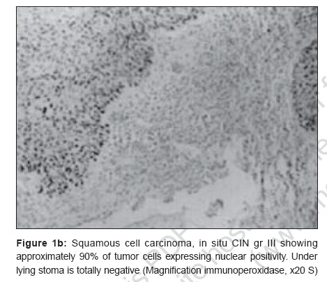
Journal of Cancer Research and Therapeutics, Vol. 3, No. 1, January-March, 2007, pp. 17-22 Original Article Implications of p53 over-expression in the outcome with radiation in head and neck cancers Lal P, Pal L, Kumar S, Dimri K, Tiwari A, Rastogi N, Singh S, Datta NR Department of Radiotherapy, Sanjay Gandhi Postgraduate Institute of Medical Sciences, Lucknow Code Number: cr07005 Abstract Background: Abnormalities in the p53 tumor suppressor gene and in the expression of its protein are commonly seen in several tumors. The prognostic implication of these p53 abnormalities was studied in 55 patients with advanced head and neck cancers.Purpose: To identify p53 as a prognostic factor in assessment of response and survival outcome to radiotherapy in head and neck malignancies. Materials and Methods: This prospective study was carried out from April 1998 to December 1999. Fifty five patients with proven squamous cell carcinoma of the head and neck region were treated by radiotherapy (RT) (n=34) with or without chemotherapy (CT) (n=21). A dose of 70Gy/35#/7 weeks was given with or without concurrent administration of weekly cisplatin (35 mg/m2). Paraffin sections obtained at the time of diagnosis, were examined immunohistochemically for p53 overexpression with monoclonal antibody DO-7 (DAKO). The scoring of p53 positive cells was carried out by a trained pathologist. Selected areas of p53 positive cells were viewed under high power field for quantitative assessment of the p53 over expression. A minimum of 1000 cells were counted and the labeling index (LI) was calculated in terms of percentage of p53 positive cells over the total number of cells counted. A 10% nuclear reactivity exhibiting chromogen positivity cutoff point was established. Observations: The data was analyzed as of January 2006. Median follow-up of all the patients was eight months (1-95 months). The median age of this study group was 58 years and of the 55 patients, 48 were males. Positive expression of p53 gene protein was documented by immunohistochemistry in 24 (44%) patients. Over expression of p53 was not associated with T or N stage, site of disease, radiation response or survival outcomes ( P =0.143). Stage was the only independent prognostic variable, both for the response to treatment (radiation) and survival ( P =0.01). Conclusions: Over expression of p53 protein, when detected immunohistochemically, does not predict for radiation response in these tumors. Keywords: p53, CHART, head and neck cancer radiation resistance Introduction p53 is a tumor suppressor gene localized to Chromosome 17 encoding 53 KD nuclear phosphoprotein and it has a principal role in controlling the cell cycle. DNA damage increases the level of p53 resulting in cell cycle arrest, DNA repair or apoptosis.[1],[2],[3] It acts as a pro-apoptotic gene, by activating BAX and BAK like molecules and caspase pathway to induce apoptosis, which is also one of the mechanisms by which radiation acts.[4] Thus, increased expression of wild type p53 should, in theory, be associated with an increase in radiosensitivity. The influence of p53 positivity on survival has been studied in tumors arising from different sites in the body. Squamous cell carcinoma of head and neck region was the subject for this study as it is a common cancer in India and p53 expression has been studied extensively in relation to treatment outcomes in different series.[1] Further, chemo radiotherapy is now considered the standard treatment in head and neck cancers in suitably selected patients as it results in organ preservation if successful. However, radio-resistance in certain tumors results in treatment failure and such patients may have better survival expectations following surgical extirpation. Thus identifying prognostic factors to radiation response is important for proper patient selection and for optimizing treatment strategies. Apart from clinical parameters, p53 is one such marker that has been linked to variable radiation response in certain in vitro and clinical studies.[1] The present study was aimed to test its predictive value in these tumors. Materials and Methods Fifty five patients of biopsy proven SCCHN cancers were inducted in this prospective study, which was carried out from April 1998 to December 1999. All patients belonged to stage III and IV with no evidence of distant metastasis. Patients who received postoperative RT were not included in this study. After clinical examination and diagnostic workup, the patients were planned for curative radiotherapy with an immobilization cast and adequate simulation. Radiotherapy was delivered by a Telecobalt (Theratron 780C) using a 3 field technique i.e., lateral parallel opposed portal for primary and upper neck and direct anterior portal for lower neck and supraclavicular region. Electron boost was given to posterior neck in case of nodes astride the spinal cord. Radical dose of radiotherapy i.e., 70Gy in 35 fractions over seven weeks was delivered in all. Thirty four out of 55 patients received RT alone, while 21 received concomitant weekly single agent cisplatin chemotherapy (@ 35 mg/m 2 ) as a part of an ongoing protocol. Cisplatin chemotherapy was administered after adequate hydration and anti-emetic cover. Statistical analysis Data including the demographic profile and p53 expression of the study group has been reported as medians and ranges. Disease-free and overall survival has been reported using the Kaplan-Meier method and the log-rank test used to determine the statistical significance of differences. P -values < 0.05 have been considered significant Results Demographic characteristics: The data was analyzed as of January 2006. The median follow-up of the present study was eight (1-95) months. The demographic features of the study population are enumerated in [Table - 1]. p53 immunostaining: In the present study 24/55 cases
(44%)
showed p53 over expression. In positive cases, the adjacent mucosa with
severe carcinoma in situ component also showed p53 overexpression [Figure
- 1]a,
b and c. Labeling index, given in [Table - 2],
was more than 50% in 58% cases. The 2 treatment protocols
did not have any difference in terms of p53 status (13 positive cases
in RT alone and 11 in CTRT arm). The response to treatment was correlated
with p53 status and is mentioned in [Table - 3].
p53 positivity and labeling score index had no bearing on the response
to treatment or outcome ( P =0.14 and 0.89 respectively). There
was no correlation between p53 and disease free (DFS) and overall survival
(OS) in this study [Figure - 2],[Figure - 3].
The Median DFS was 5 and 0 months in the p53 positive and negative arms
respectively ( P =ns). Similarly, the median OS was 8 and 7 months
in the p53 positive and negative arms respectively ( P =ns). Discussion p53 mutations occur commonly in head and neck cancer patients. Various studies have shown p53 positivity in squamous cell carcinoma of the head and neck region in 33-73% of the cases.[2],[3] The wide range of positive values could have been due to differences in the staining techniques used. The present study has shown p53 gene protein expression in 44% of these tumors. Detection of mutant p53 protein can be carried out by either studying the DNA sequencing or studying the region and type of mutation; or by detecting the mutant p53 protein within the tumor tissue. They have been studied extensively by various methods, of which IHC is one of them. The wild type p53 has very short half-life (20 minutes) and thus not detected by IHC. Mutant p53 has longer half-life and can thus be detected immunohistochemically. Immunohistochemistry for the analysis of p53 has been criticized, partly because of conflicting results in clinical studies. The method does not necessarily detect all mutations.[5] This also includes the pitfalls of IHC- for instance DO 7 clone picks up both wild and mutant type p53. In the present study as well, DO7 monoclonal antibody was used, which can recognize both wild and mutant p53 protein. Heterogeneity of labeling may occur because not all tumor cells harbor mutant p53 gene and not all p53 mutations result in accumulation of p53 protein. For instance, Nylander et al showed 50% immuno-negativity tumor to have p53 mutations. They concluded that the absence of p53 expression does not necessarily indicate that p53 mutation is not present.[6] Others also suggest that different antigen retrieval techniques may also be responsible for obscuring any correlation by creating falsely high positive cases.[7] Raybaud-Diogene et al published their results of a large clinical trial, which has reported a significant influence of p53 on local control of disease in patients with SCC of head and neck region.[1] They encountered 49% positivity in these tumors. Amongst the markers studied, p53 was the strongest factor for assessing the primary radiation response. The authors observed that p53 positivity and Ki67 labeling index together were documented in poor responders to radiation. The evidence of association between p53 and local tumor control probability has been equivocal. On one hand Koch et al and Watling et al suggest an association between aggressiveness or lack of responsiveness in p53 positive tumors[8],[9] and on the hand other authors do not.[7],[10],[11] The reason why some workers did not obtain positive results was possibly because they were small studies with heterogenous population. Regarding the influence of host and tumor factors such as age, gender and primary site, no such association was seen with p53 positivity in the present study. This is unlike what has been shown in some of the other studies.[12] A study reported by Frank et al showed strong p53 positivity in younger patients and advanced stage of presentation.[12] Amongst the treatment-related factors, there is ample evidence that concurrent chemo radiotherapy is the standard of care in head and neck cancers.[13] Since these patients were a part of ongoing trial during that time period comparing chemo radiotherapy with RT alone, this subset of patient failed to show the benefit with addition of chemotherapy. This was possibly due to disbalance between the two arms in this subset i.e., CTRT arm patient had higher nodal burden (N3 - 19% and 9% respectively) and stage IV disease as compared to RT alone (60% and 71% respectively). Similarly, comparison between the p53 positive and negative within the protocols did not show any difference in survival. Various workers have studied the prognostic significance of p53 in a particular subsite of head and neck cancers. For instance, Koelbl assessed p53 status in oral lesions but did not find any correlation.[14] Similarly, other authors studied the influence of p53 status in base tongue, pharyngeal wall and hypopharynx, larynx.[15],[16],[17] Sauter et al found improvement in survival in patients whose base tongue cancers abnormally expressed p53.[15] Lassaletta studied the p53 expression in pharyngeal wall tumors treated with induction chemotherapy. They found that overall p53 positivity rate was 73% and response rates to chemotherapy tended to be higher in the p53 negative group ( P =0.07). Similarly, the risk of recurrence was lower in the p53 negative group.[16] The present study, although a heterogenous group of head and neck cancers, showed no prognostic bearing of p53 with the radiation response, DFS and OS. Most studies with similar patient data report similar lack of correlation. Wilson et al expected to define the role of p53 in radiation sensitivity in an accelerated schedule of CHART. Acceleration overcomes one confounding factor i.e. proliferation. However, correlation of p53 as a marker with DNA damage and response could not be made. They attempted to semiquantitate two aspects of heterogeneity- the staining pattern and intensity of staining. Only the staining intensity had association with clinical or biological characteristics.[18] Koelbl et al also do not regard p53 status as parameter for radiosensitivity. In their series the most important prognostic factor reported for control and survival was degree of tumor regression.[14] Awwad et al felt that p53 was not predictive of patients'poor survival or freedom from disease. This multivariate analysis revealed stage as the most significant prognostic factor.[9] In the present study too, stage emerged to be an independent prognostic variable when all stages were compared, but it lost its significance once advanced stages i.e., III and IV only were considered. Oral lesions fared worse, irrespective of p53 status, but since the number is small, it was difficult to draw any conclusions. Certain authors have questioned whether local control rate after combined treatment is the clinical manifestation of high radiosensitivity caused by the p53 status.[14] Others felt that accumulation of p53 protein does not predict survival although it may predict organ conservation.[10],[19] Narayana et al studied recurrent T1 glottis and observed 82% p53 over expression rate compared to 29% in the control group (not recurred) ( P < 0.0001). High intensity of p53 was seen in recurrent cases. They chose early glottis so that only the early mutations such as p53 mutations would be present. In advanced disease, several genetic changes may have occurred which mask the true influence of p53. This study showed that p53 was an important prognostic factor and was independent of histological grade.[17] Similarly, Jackel et al in their multivariate analysis of 88 primary laryngeal squamous cell carcinoma found p53 to be of independent prognostic significance unlike BCL-2.[20] The possible reason for lack of correlation, in the present study and others, could be that carcinogenesis is a multifactorial process involving multiple mutations, of which p53 is one of the earlier one. So, since p53 alone is not responsible for response to radiation, its detection along with other biological markers may be a reliable method of prediction. Additionally, without comparison with surgical series, it is difficult to determine whether poor outcome of p53 positive cases is due to radioresistance or intrinsic biologic behavior. Conclusion The findings of the present study and from the review of literature, it appears that p53 is only one of the components regulating the cell cycle. Moreover it is unlikely that radioresistance would be influenced by a single event such as mutation of p53 oncogene. Hence, possibly several biological markers need to be studied along with p53 to predict the radiation response. References
Copyright 2007 - Journal of Cancer Research and Therapeutics The following images related to this document are available:Photo images[cr07005f2.jpg] [cr07005f1c.jpg] [cr07005f3.jpg] [cr07005f4.jpg] [cr07005t2.jpg] [cr07005f1a.jpg] [cr07005f5.jpg] [cr07005t4.jpg] [cr07005t3.jpg] [cr07005f1b.jpg] [cr07005t1.jpg] |
| |||||||||

![[Table - 1]](/showimage?cr/photo/cr07005t1.jpg)
![[Figure - 1]a](/showimage?cr/photo/cr07005f1a.jpg)
![[Table - 2]](/showimage?cr/photo/cr07005t2.jpg)
![[Table - 3]](/showimage?cr/photo/cr07005t3.jpg)
![[Figure - 2]](/showimage?cr/photo/cr07005f2.jpg)
![[Figure - 3]](/showimage?cr/photo/cr07005f3.jpg)
![[Table - 4]](/showimage?cr/photo/cr07005t4.jpg)
![[Figure - 4]](/showimage?cr/photo/cr07005f4.jpg)
![[Figure - 5]](/showimage?cr/photo/cr07005f5.jpg)