 |
| About Bioline | All Journals | Testimonials | Membership | News |
|
||||||
|
||||||
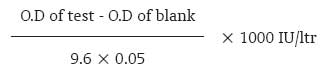
Journal of Cancer Research and Therapeutics, Vol. 3, No. 3, July-September, 2007, pp. 167-168 Brief Communication Serum total glutathione-s-transferase levels in oral cancer Prabhu Krishnananda, Bhat GopalakrishnaP Department of Biochemistry, Kasturba Medical College, Manipal - 576 104, Karnataka Code Number: cr07043 Abstract We conducted a study wherein serum total glutathione-s-transferase levels were measured in patients (n = 27) with various stages of biopsy proven oral cancer (squamous cell carcinoma) and age and sex matched healthy human volunteers (n=10). In all patients with oral cancer, serum total glutathione-s-transferase was measured before the onset of treatment. There was a significant increase in serum total glutathione-s-transferse levels in patients with stage IV oral cancer as compared to stage II (P = 0.001) and stage III (P = 0.002) oral cancer. This shows that alterations in serum total Glutathione-s-transferase levels may have a role in cancer progression.Keywords: Glutathione-s-transferase, oral cancer, progression Introduction Oral cancer accounts for about 8% of all malignant growths. Men particularly over 40 years are affected twice as often as women. The exact cause is unclear but smoking, alcohol, tobacco chewing, poor oral hygiene, chronic irritation etc are implicated. Reactive oxygen species (ROS) like superoxide, hydrogen peroxide etc. have been implicated in many diseases including cancer. ROS have been known to play an important role in initiation and progression of multi-step carcinogenesis. [1] Alterations in circulating antioxidants and free radical scavengers like glutathione-s-trnasferase (GST) have been linked with various epithelial malignancies including oral cancer. [1],[2],[3],[4],[5],[6],[7],[8],[9] In this study, we evaluated the levels of serum total GST in oral cancer patients and healthy controls.Materials Reduced glutathione C 10H17O 6 S and 1-chloro-2, 4, dinitro benzene (CDNB) were purchased from Sigma Chemical Company. All other reagents used were of Reagent grade. Deionized water was used throughout the study. Permission for the study was granted by the Institutional Ethics Committee. Informed consent was taken from controls (healthy volunteers, n = 10) and patients (n = 27). The age of the subjects (n =37) was 56.4±1.67 yrs [Table - 1]. All the patients were admitted for radiotherapy. In cases blood was withdrawn just before initiation of treatment. Serum total GST levels were measured in biopsy proven cases of oral cancer (n=27) with biopsy showing moderate to well differentiated squamous cell carcinoma. Methods Serum GST was estimated by CDNB method. [10],[11],[12]Reagents a. Phosphate Buffer: 0.1M, pH-6.5 prepared with deionized water and stored in brown bottle in refrigerator. Assay GST was estimated in 1ml of incubation mixture containing 850 µl of 0.1 M phosphate buffer pH 6.5 and CDNB reagent (20 mM) 50 µl, preincubated at 37°C for 10 min. Reaction was started by adding 50 µl of 20 mM GSH and 50 µl of serum. Reaction was followed at 1 min interval for 5 min by measuring absorption at 340 nm. Simultaneously, blank was run by substituting deoinized water for serum. Then O.D change/min was calculated. GST was estimated by using the molar extinction coefficient [9.6 mM -1 cm -1 ] of GST. [11] Formula
Statistical analysis Kruskal Wallis test was used to analyze the results and it showed a significant difference ( P = 0.001). In case of significant difference, pair-wise comparison between control and various stages were done by Mann- Whitney Test adjusting a for the number of pairs to be compared. (significance at the level: 0.05/6 = 0.0083) Results and Discussion Comparison of serum total GST between control and cancer patients using Kruskal Wallis showed a significant difference ( P = 0.001). However, the paired comparison between control and various groups by Mann Whitney did not show a significant difference. ROS are tumorogenic by virtue of their ability to increase cell proliferation, survival, cellular migration and also by inducing DNA damage leading to genetic lesions that initiate tumorogenicity and sustain subsequent tumor progression. As shown by earlier studies, loss of antioxidant capacity of cell in early dysplasias can trigger initiation and progression of cancer. [13] Our results showed a decrease in serum total GST in early cancer than the control [Table - 2] which may have triggered the initiation and progression of cancer. Many studies also showed progressive increase of GST with advancing cancer and has been associated with poor prognosis and development of drug resistance. [14],[15] In our study there was an increase in serum total GST in later stages of cancer. This enhanced antioxidant capacity made the tumor tissues less susceptible to oxidative stress conferring specific growth advantage. [16] References
Copyright 2007 - Journal of Cancer Research and Therapeutics The following images related to this document are available:Photo images[cr07043t1.jpg] [cr07043t2.jpg] |
| |||||||||

![[Table - 1]](/showimage?cr/photo/cr07043t1.jpg)
![[Table - 2]](/showimage?cr/photo/cr07043t2.jpg)