 |
| About Bioline | All Journals | Testimonials | Membership | News |
|
||||||
|
||||||
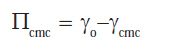
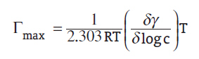
Journal of Cancer Research and Therapeutics, Vol. 3, No. 4, October-December, 2007, pp. 198-206 Original Article Surface and antitumor activity of some novel metal-based cationic surfactants Badawi AM, Mohamed MekawiAS, Mohamed MZ, Khowdairy MM Surfactant Laboratory, Petrochemicals Department, Egyptian Petroleum Research Institute, Nasr city, Cairo Code Number: cr07051 Abstract The development of anticancer metal-based drugs was attempted by reacting dodecyl amine with selenious acid to produce a quaternary ammonium salt which was then converted to copper and cobalt cationic complexes via complexing the first compounds with copper (II) or cobalt (II) ions. The surface properties of these surfactants were investigated. The surface properties studied included critical micelle concentration (CMC), maximum surface excess (Γmax ), and minimum surface area (Amin ). Free energy of micellization (∆G o mic ) and adsorption (∆Go ads ) were calculated. Antitumor activity was tested by using Ehrlich ascites carcinoma (EAC) as a model system of mice cell tumor. The compounds were also tested in vitro on five human monolayer tumor cell lines: MCF 7 (breast carcinoma), HEPG 2 (liver carcinoma), U 251 (brain tumor), HCT116 (colon carcinoma), and H460 (lung carcinoma). FTIR spectra, elemental analysis, and H 1 NMR spectra were performed to insure the purity of the prepared compounds.Keywords: Antitumor activity, cationic surfactant, critical micelle concentration Introduction In terms of abundance, selenium (Se) ranks 70 th among the elements and constitutes approximately 10-5 % of the Earth′s crust. [1] Se compounds are known to act as anticancer agents, both in intact animals and in cellular systems. [2],[3] Se induces cell cycle arrest at different phases, depending on the chemical form of Se and the cell type. [4],[5] The inhibitory effect on cell proliferation, with a preference for tumor cells vs nontransformed cells, is considered to be a mechanism for the anticarcinogenic capability of Se. [6] Se-induced apoptosis in cancer cells was related to its chemopreventive activity. [7] Several groups have shown that selenocompounds induce apoptosis in cell culture systems. [8],[9] The chemopreventive action of Se compounds have been suggested to result from inhibitory effects on carcinogen activation and the potentiation of the immune system. [10] In addition, compounds such as selenite induce a number of cytotoxic effects in tumor cells in vitro , [11] which suggests that this may be one of the mechanisms involved in the inhibition of tumor development in vivo . Organometallic amphiphiles are a new class of surfactants. Their various novel chemical and physical properties of adsorption and aggregation, such as selective binding, active control of surface tension, cytotoxic activity, and charge transfer, have been widely investigated in recent years. Studies on the chemistry of transition metal complex surfactants have received a sustained high level of attention due to their relevance in various redox processes in biological systems; they are considered to be promising agents for the development of new antitumor drugs. The main goal of cancer therapy is to attain the maximum therapeutic damage of tumor cells with the minimum concentration of the drug. This can be achieved, in principle, via selective antitumor preparations, the cytostatic effects of which would be restricted within tumor tissue. While 100% selectivity may be impractical, achievement of reasonably high selectivity seems to be a feasible aim. The bioenergetic status in tumor was selective and affected by the metal complexes. Minimization of signals of high-energy phosphate was observed after injection of the complexes. An increase in the number of DNA single-strand breaks registered in tumor tissue, supporting the suggestion that the complexes may directly affect DNA; however, the action of these complexes as antitumor agents was found to be dependent on the type of tumor cell line tested. [12] Synthetic Procedure Synthesis of dodecylammoniumhydrogen selenites Stoichiometric amounts of selenious acid were mixed with dodecylamine at room temperature in ethyl alcohol and then stirred until the precipitation stopped. The (white yellowish, reddish, and pink) precipitant was filtered, washed by ethyl alcohol, and then recrystallized by diethyl ether. [13] The products are designated as II g and have the general formula: RN+H 3 HSeO 3 where R = dodecyl. Synthesis of metal complexes Synthesis of cobalt (II) hydrogen selenite dihydrate Hydrogen selenite cobalt (II) dehydrate, with the formula Co(HSeO 3 ) 2 .2H 2 O, is a new biologically active chemical compound that is used in alga biotechnology for obtaining biomass blue-green microalga of spirulina. It has a high content of organic-combined Se, which may serve as a biological raw material for obtaining Se-containing medical preparations (drug). For obtaining cobalt (II) hydrogen selenite dihydrate, selenious acid (H 2 SeO 3 ) is reacted with basic cobalt (II) carbonate [Co(OH 2 ) 2 CO 3 ], which is prepared by mixing aqueous solutions of equimolar amounts of CoCl 2 and Na 2 CO 3 . The precipitate is washed till the absence of foreign ions. 2H 2 O + Co Cl 2 + Na 2 CO 3 → CoCO 3 .2H 2 O + 2NaCl An aqueous solution of 2 gm (0.016 mol) H 2 SeO 3 in 10 ml water is added to a warm solution of the freshly prepared Co carbonate 1.22 gm (0.008 mol) in 10 ml water. The obtained solution is filtered and kept at room temperature for crystallization for 2 days. Crystalline prisms of red color are formed. The crystals are filtered, washed with water, and dried in air. [14] Co (HSeO 3 ) 2 has a molecular weight of 350.892 gm/mol. Co (II) hydrogen selenite dihydrate occurs as prismatic crystals of red color. It is stable in air and soluble in water and alcohol. With respect to free water and thermogravimetric properties, the temperature of removal of 2 molecules of water is 100-110°C. 100°C Synthesis of copper (II) hydrogen selenite dihydrate For obtaining copper (II) hydrogen selenite dihydrate, selenious acid (H 2 SeO 3 ) is reacted with basic copper (II) carbonate [Cu(OH 2 ) 2 CO 3 ], which is prepared by mixing aqueous solutions and equimolar amounts of CuCl 2 and Na 2 CO 3 . The precipitate is washed till the absence of foreign ions. An aqueous solution of 2 gm H 2 SeO 3 in 10 ml water is added to a warm solution of freshly prepared Cu carbonate 1.28 gm in 10 ml water. The obtained solution is filtered and kept at room temperature for crystallization for 24 h, when crystalline prisms of blue color are formed. The temperature of removal of 2 molecules of water is 100-110°C. Synthesis of copper and cobalt ammonium hydrogen selenite complexes Cobalt or copper fatty dodecylammonium hydrogen selenite complexes were prepared by refluxing two moles of dodecylammonium hydrogen selenite (II g ) with one mole of cobalt or copper hydrogen selenite in ethyl alcohol for 2 h. The products were designated as (II h,i ). 2RN+H 3 (HSeO 3 ) - + M (HSeO 3 ) 2 → [RNH 3 ]+ 2 M [HSeO 3 ] - 4 The products were purified and recrystallized three times in petroleum ether and then washed with diethyl ether. The products were kept in desiccators until use. The general formula for the metal complexes is as follows: [RN+H 3 ] 2 [M (HSeO 3 ) 4 ] -2 where R = dodecyl and, M: Co+2 or Cu+2 . Methods of Analysis and Instruments Infrared spectra for the prepared surfactants were measured using Avatar 230 FTIR spectrophotometer to measure the intensity of the absorption bands for the prepared surfactants. The measurements were carried out at the Egyptian Petroleum Research Institute. The elemental analysis for the obtained surfactants was carried out using Elemental Analyzer, Model: Vario elementar. The measurements were carried out at the Micro Analytical Center, Faculty of Science, Cairo University. Proton nuclear magnetic resonance measurements (H 1NMR) were performed on a Varian-Gemini-200 instrument and the samples were run in deuterated chloroform (CDC13), Cambridge Isotope Laboratories) at the Micro Analytical Center, Faculty of Science, Cairo University. Atomic absorption spectrometer (AAS) measurements for copper and cobalt analyses were performed with AAS (flame absorption) PerkinElmer; the detection limits for this analysis are 0.005 gm/20 ml for copper and 0.003 gm/20 ml for cobalt. The analysis was done at the Micro Analytical Center, Faculty of Science, Cairo University. Evaluation Methods of Surface Active Properties Surface and interfacial tension measurements Surface and interfacial tension measurements of the prepared surfactants were made at (25°C) with Du Nouy tensiometer (Kruss type 8451), using distilled water solution of 0.1% weight concentration. [15] The surface tension of the used distilled water was 73 mN/m and the interfacial tension between medicinal paraffin oil and distilled water was 56.2 mN/m. Surfactant solutions were aged for ½ h before any measurements were made. Three readings were made on each sample to determine any change with time and to obtain an average value. [16] Emulsifying power Emulsifying power or emulsifying time (in seconds) was determined by mixing surfactant solution (0.1 gm/10 ml) and paraffin oil (10 ml) in a measuring cylinder with vigorous shaking (10 times); the tube was allowed to stand till any separation of the two phases appeared. [17],[18] Efficiency (PC 20 ) The efficiency (PC 20 ) was determined as the concentration (mol/l) capable of suppressing the surface tension by 20 dyne/cm. [19] The efficiency was determined by extrapolating from γ = 52 to the linear portion before CMC of the γ vs Log C plot [20] at 25°C. Effectiveness (ІІcmc ) The surface tension (γcmc ) values at CMC were used to calculate the values of surface pressure (effectiveness), using the following expression: Where γo is the surface tension measured for pure water at the appropriate temperature and γcmc is the surface tension at CMC. The effectiveness of adsorption is an important factor that determines such properties of the surfactant as foaming, wetting, and emulsification, since tightly packed, coherent interfacial films have very different interfacial properties from loosely packed, noncoherent films. [21] Determination of CMC CMC of the prepared surfactant was determined by the surface tension method. [22] In this method, values of the surface tension obtained for various concentrations of aqueous solutions of the prepared surfactants were plotted vs the corresponding concentrations. Maximum surface excess (Γmax ) The surface excess concentration is defined as the surface concentration at surface saturation; the maximum surface excess (Γmax ) is a useful measure of the effectiveness of adsorption of the surfactant at the water-air interface, since it is the maximum value to which adsorption can attain.
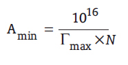
where R = 8.314 Jmol-1 K-1 , T is absolute temperature, (δγ/δ log C) is the slope of the γ vs Log C plot at 25°C. [23] A substance which lowers the surface tension is thus present in excess at or near the surface, i.e., when the surface tension decreases with increasing the activity of the surfactant, Γ is positive Minimum surface area (A min ) A min is the minimum area per molecule of the prepared compounds at the interface and is calculated from the following equation:
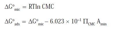
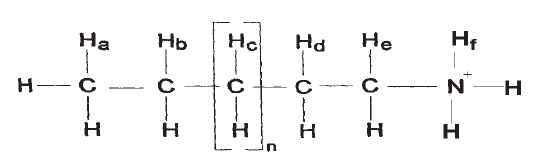
where N is Avogadro′s number and Γmax is the maximum surface excess. The standard free energies of micellization ∆G o mic and adsorption ∆G o ads Understanding the process of micellization and adsorption are important for explaining the effects of structural and environmental factors on the value of the CMC and for predicting the effects on it of new structural and environmental variations. Standard free energy of micellization (∆G o mic ) and adsorption (∆G o ads ) play an important role in facilitating such understanding. The standard free energy of micellization and adsorption are given by: The interfacial activity (І activ.) І activ. is expressed by physicochemical parameter ∆G ads /A min , where ∆G ads is the standard free energy of adsorption of the surfactant at the air-solution interface and A min is the minimum cross-sectional area of the surfactant. Antitumor activity of the Erlisch ascites carcinoma (EAC) [24] A set of sterile test tubes were used, in which 2.5 x 10 5 tumor cells per ml were suspended in phosphate buffer saline. Then 25, 50, 100 µg/ml of the drug were added to the suspension, which was then kept at 37°C for 2 h. Trypan blue dye exclusion test was then carried out to calculate the percentage of nonviable cells. Potential cytotoxicity measurements by SRB assay Potential cytotoxicity of the compound(s) was tested using the method of Skehan et al . [25] The tumor cell lines - MCF 7 (breast carcinoma), HEPG 2 (liver carcinoma), U 251 (brain tumor), HCT 116 (colon carcinoma), and H 460 (lung carcinoma) - 10 4 cells/well, had been supplied from National Cancer Institute, Cairo, Egypt; they were plated in 96-multiwell plates for 24 h before treatment with the compound(s) to allow attachment of the cells to the wall of the plate. Different concentrations of the compound under test (0, 1, 2.5, 5, and 10 µg/ml) were added to the cell- monolayer. Triplicate wells were prepared for each individual dose. Monolayer cells were incubated with the compound(s) for 48 h at 37°C in an atmosphere of 5% CO 2 . After 48 h, the cells were fixed, washed, and stained with sulforhodamine B stain. Excess stain was washed off with acetic acid and the attached stain was recovered with Tris-EDTA buffer. Color intensity was measured in an ELISA reader. The relationship between the surviving fraction and the drug concentration was plotted to get the survival curve of each tumor cell line after the specific compound was added. Results of FTIR Data The chemical structures for surfactant samples were accorded in FT-IR [Table - 1]. The FT-IR absorption spectra showed an absorption band at the 2365-2377 cm-1 region, indicating that the primary amine band was disappearing by emerging ammonium ion (R N+ H 3 ). In addition, there was a strong band at 719-762 cm-1 , indicating the presence of multiple (CH 2 ) groups. The very strong bands at the 2849-2855 cm-1 region for all the prepared compounds were due mainly to the methyl symmetric stretching vibration. The sharp band at 2921-2966 cm-1 was observed for all prepared compounds due to the stretching vibration of the symmetric methylene group. In case of amine complexes, a new band was shown at 571-580 cm-1 , which was evidence for the presence the cobalt or copper ligand, alike with the stretching band of CO-N (DMG dimetheylglyoximate) was assigned at 512 cm-1 . [26] The results is generally in agreement with the expected correlations. H1NMR structural studies The H1NMR spectrum for some selected cationic surfactants are represented as shown in the following example:
The first peak shows the methyl protons (H a ) with two neighboring protons, appearing at δ = 0.84 ppm, while the second peak shows the methylene protons (H b ) with five neighboring protons appearing at 1.5 ppm. The third peak shows methylene protons (H c ) n with four neighboring protons appearing at δ = 1.23 ppm. The fourth peak shows methylene protons (H d ) with four neighboring protons; this signal is shifted to up field because of the electronegativity of the nitrogen atom and therefore appears at δ = 2.5-2.7 ppm. The fifth peak shows methylene protons (H e ) with five neighboring protons appearing at δ = 3.3-4.364 ppm. The sixth peak shows amine protons (H f ) with five neighboring protons appearing at δ = 7.5-7.7 ppm. It is shifted to up field due to the positive charge on the nitrogen atom. In conclusion, the spectra for all prepared compounds show the same seven main characteristic peaks. The difference between these compounds is in the intensity of the methylene proton signal and that of the nitrogen atom. The intensity of these signals increases due to the doubling of the methylene groups and ammonium ions in case of complexes. Elemental analysis data A further structural confirmation of the prepared surfactants was given by elemental analyses. The data of elemental analysis are presented in [Table - 2]. The data indicate that the calculated percentages of C, H and N are close to the found measurements. From [Table - 2] the carbon percent conformity were found running smoothly from 98.5% to 101%, while hydrogen, nitrogen, and chloride were running slightly higher than the calculated value. This was considered as an acceptable and tolerated value. Atomic absorption spectrometer data AAS results in [Table - 3] confirmed the prepared complex compounds, since there is no significant error percent between the expected and the experimental values. Surface Properties of the Prepared Cationic Surfactants Surface properties of the prepared cationic surfactants were measured and are tabulated in [Table - 4]. Surface and interfacial tensions Surface tension is a characteristic property of liquids. The phenomenon is due to the attraction force between the molecules at the surface. The surface tension value of distilled water at 25°C was found to be 72 mN/m and is attributed to the attraction forces between water molecules at the water surface due to the hydrogen bonds. If any foreign molecules are present at the water surface there is a disturbance in the forces, leading to a decrease in the surface tension. Surfactant molecules tend to be adsorbed at the air-water interface at lower concentrations. Hence, by increasing the surfactant concentration the surface tension of the resulting solution can be decreased gradually. Surface and interfacial tensions of the prepared cationic surfactants and their corresponding metal complexes were measured and are shown in [Table - 4]. The surface tension values of the cationic complexes (II h,i ) were found to be lower than that of their parent cationic surfactant. That could be due to the increase in the hydrophobicity of these complexes as compared to that of the parent cationics, which is due to the presence of two ligands coordinated to the metal ion form giant structure of the complex containing higher number of methylene groups, which increases the water-surfactant molecule interactions, forcing them to the air-water interface. [27] Hence, the surface tension is depressed considerably [Table - 4]. In fact, these results suggest that two alkyl chains in one molecule linked by a metal ion enhances the adsorption and aggregation properties by strengthening the inter- or intra-molecular hydrophobic interaction. [28] The uniqueness of the metal-surfactant coordination complexes lies in the fact that the bond between the head group and the tail part of the surfactant is a coordinate bond and the surfactant contains a higher charge on the head group, which leads to more repulsion in the bulk of the aqueous solution and increasing adsorption onto the surface. This may also be attributed to a difference in the steric packing of the complexes, which causes a different arrangement of the alkyl chains on the surface. [29] Emulsifying power The emulsifying power of the prepared surfactants is listed in [Table - 4] as a function of time. It is clear from the data that all the prepared surfactants show adequate emulsifying power towards paraffin oil. CMC of the prepared surfactants Surfactants form aggregates of molecules or ions called micelles, which are formed when the concentration of the surfactant solute in the bulk of the solution exceeds a limiting value, the so-called critical micelle concentration (CMC), which is a fundamental characteristic of each solute-solvent system. If the properties of a surfactant solution are plotted as a function of the concentration of the surfactant, the properties usually vary linearly with the concentration up to the CMC, at which point there is a break in the curve as shown in [Figure - 1]. The results in [Table - 5] and [Figure - 1] show that on complexing the cationic surfactants with cobalt or copper ions, marked depression is observed in the CMC values compared to that of the parent cationics. This could be explained by the unique property of the metal complexes in water, i.e., the complexes retain their unity in the solutions, which increases their volume in the aqueous media; repulsion then occurs between the hydrophobic chain and the water molecules. This repulsion facilitates two processes at the same time, i.e.:
The application of these molecules in commercial processes would allow us to dramatically reduce the concentration of surface active material used, while maintaining the same level of performance. Thus, it is concluded that these metal surfactant complexes have more ability to associate themselves, forming aggregates, compared to the ordinary synthetic quaternary ammonium salts, which suggests that the introduction of a metal ion into the hydrophilic part of the amphiphile can remarkably enhance the ability to aggregate. Effectiveness (ІІcmc) The effectiveness (ІІcmc ) is determined by the difference between surface tension values at CMC (γcmc ) and the surface tension measured for pure water at the appropriate temperature (γo ). The most efficient one is that which gives the greatest lowering of surface tension for a given CMC. (III i ) was found to be the most efficient one [Table - 5] because it achieved the maximum reduction of the surface tension at CMC [Figure - 1]. Efficiency (PC 20) The efficiency (PC 20 ) was determined by the concentration (mol/l) capable of suppressing the surface tension by 20 dyne/cm. Values of efficiencies of the prepared surfactants are shown in [Table - 5]. The efficiency increases with increasing molar ratio of methylene units. This is due to the fact that the efficiency of adsorption at interfaces increases linearly with increase in the carbon atoms in the hydrophobic group. [20] Maximum surface excess (Γmax) The number of surfactant molecules at the air-water interface at the CMC at 25°C is expressed by Γmax . A substance lowering the surface energy is thus present in excess at or near the surface, i.e., when the surface tension decreases with increasing activity of surfactant, Γmax is positive. It is evident from [Table - 5], in case of prepared parent cationic surfactants, that by increasing the number methylene units Γmax increases. Complexing the cationic surfactant with cobalt and copper ions contributes to the migration of molecules to the water-air interface, causing a consequent increase in Γmax values. Minimum area per molecule (A min) The minimum surface area is defined as the area occupied by surfactant molecules at the air-water interface when the solution is at equilibrium. The results given in [Table - 5] indicate that the consequent increase of Γmax leads to crowding at the interface, which causes a decrease in A min values. This is because the minimum surface area decreases with increases in the hydrophobic chain length of the synthesized surfactant molecules. The data on the minimum surface area of the short chain surfactant molecules showed higher A min of these molecules compared to the longer hydrophobic chains The standard free energies of micellization (∆G o mic ) and adsorption (∆G o ads) From [Table - 5] it can be seen that the values of ∆G o mic and ∆G o ads are always negative, indicating the spontaneousness of these two processes, but there is more increase in negativity of ∆G o ads than of ∆G o mic , indicating the tendency of the molecules to be adsorbed at the interface. Antitumor action of the prepared compounds Dodecylammonium hydrogen selenite with its cobalt and copper complexes were investigated as potential selective anticancer prodrugs. They were tested by using Ehrlich ascites carcinoma (EAC) as a model system of mice cell tumor. These compounds were also tested in vitro on five human monolayer tumor cell lines: MCF 7 (breast carcinoma), HEPG 2 (liver carcinoma), U 251 (brain tumor), HCT 116 (colon carcinoma), and H 460 (lung carcinoma). Evaluation of antitumor activity of the EAC The choice of EAC cells as a model system was based on the finding that it is an excellent tool for studying the biological behavior of malignant tumors and drug action within cells. [30] The line of EAC which was used in the present study was kindly supplied by the National Cancer Institute, Cairo, Egypt, and was maintained in female Swiss albino mice through weekly IMP transplantation of 2.5 x 10 6 tumor cells/mouse. EAC cells were obtained by needle aspiration under aseptic conditions. The ascitic fluid was diluted with sterile saline so that 0.1 ml contained 2.5 x 10 6 cells when counted under a microscope using a hemocytometer. In vitro antitumor activity of these compounds was determined according to the percentage of nonviable cells (NVC %), which was calculated by the following equation: NVC% = [number of NVC/total number of cells] x 100 The results of these experiments are summarized in [Table - 6]. As shown in [Table - 6], increasing the concentration of dedocylammonium hydrogen selenite (II g ) and its cobalt (II h ) or copper (II i ) complexes in the EAC media was accompanied by progressive increase in the NVC %. This is due to the fact that by increasing the concentration of cationic surfactant the adsorption of ions on cell membranes increases, leading to increase in penetration and antitumor activity. The inhibition of cell viability percent showed that the II h (cobalt complex) is the most active one at a concentration of 100 µg/ml, the NVC % reaching up to 100%. This means that the drug at this concentration causes the death of all the tumor cells, while at concentration 50 µg/ml the percentage reached 80%. At a concentration of 25 µg/ml the NVC % reached 50%. With II i (copper complex), at a concentration of 100 µg/ml the NVC % reached 80% and at a concentration of 50 µg/ml it reached 60%. From these results, II h is seen to be the most active of all the derivatives; cobalt complexes seem to offer promise due to the high electron affinity of the metal (which increases its ability to bind DNA) and the ready reducibility of the compounds. [31] It can also be seen that II g has the least toxic effect of all the derivatives on EAC cells. Evaluation of cytotoxic activity on human tumor cell lines The main goal of cancer therapy is to attain the maximum therapeutic damage of tumor cells using the minimum concentration of the drug. This can be achieved, in principle, via selective antitumor preparations, the cytostatic effects of which would be restricted within tumor tissue. While 100% selectivity may be impractical, achievement of reasonably high selectivity seems to be a feasible aim. The results of the cytotoxic activity on human tumor cell lines was determined according to the dose values of drug exposure required to reduce survival in the cell lines to 50% (IC 50 ). The experimental results are recorded in [Table - 7] and plots of surviving fraction vs concentration in micrograms are shown in [Figure - 2],[Figure - 3],[Figure - 4],[Figure - 5],[Figure - 6]. From the results recorded in [Table - 7] and [Figure - 2],[Figure - 3],[Figure - 4],[Figure - 5],[Figure - 6], some of the compounds tested exhibited high activity in vitro system on the tumor cell line investigated. II h have the highest cytotoxic effect on H460 , MCF7, and HCT116 ; the doses at which the survival was reduced to 50% (IC50 ) in each of these cell lines was 1.1, 9.6, and 8.7 µg/ml, respectively Respectively as shown in [Figure - 2],[Figure - 3],[Figure - 6]. Also, IIh showed good cytotoxic activity on HEPG 2 (IC50 = 8.63 µg/ml), as shown in [Figure - 4]. It should be noted that the action of these compounds as antitumor agents is found to be dependent on the type of tumor cell line tested but, as shown from the results, IIh (cobalt complexes) show excellent cytotoxic activity against several tumor cell lines and, at very low concentrations, reduces the survival to 50%. This is due to the fact that cobalt complexes have a capacity to reduce the energy status in tumors as well as to enhance tumor hypoxia, which also influences their antitumor activities. It may be also concluded that the level of cellular damage inflicted by these complexes depends on the nature of their axial ligands. There is evidence that cobalt complexes cause significant changes in metabolism, namely activation of lipid peroxidation, DNA damage, and reduction of the bioenergetic status of tumor tissues. In general, the high selectivity of action by redox-active cobalt complexes upon tumors is due to their specific reactivity .[12] While, as shown in [Figure - 4],[Figure - 5], IIi has the highest cytotoxic effect on HEPG 2 and U251 (IC50 = 8.39 and7.83 µg/ml), it also shows good cytotoxic activity on HCT116 (IC50 = 8.93 µg/ml) as shown in [Figure - 6]. This may be due to oxidative reaction of Cu 2+ and site-directed mutagenesis of the putative catalytic base inhibiting both serine and tyrosine protein kinase activity, suggesting that one active site is involved in both activities, which leads to suppression of the resistance of tumor cells against cytotoxic agents. [32] Yang et al . reported the ability of copper complexes to inhibit DNA synthesis of tumor cells, which is closely related to the antitumor mechanism of the complex. [33] Copper complexes exhibit superoxide dismutase-like activity (superoxide dismutase is used as an anti-inflammatory agent and as lipid soluble). This property enables the compound to penetrate cell membranes and become inter-cellular. [34] Finally, in our research, we found that copper and cobalt complex surfactants affect tumor tissue at very low concentrations - at values lower than their CMC values - which means that there is a strong relationship between the very small values of CMC of these compounds and the ability to reach IC50 values under very low concentrations. This is due to the fact that increasing the concentration of cationic surfactant causes an increase in the adsorption process on cell membranes till the CMC is reached; after this the adsorption slowly decreases and then stops due to the formation of micelles, which prevents mobility and suppresses antitumor activity. Dedoceyl ammonium hydrogen selenite does not reach to IC50 for any of the tested human monolayer tumor cell lines as shown in [Figure - 2],[Figure - 3],[Figure - 4],[Figure - 5],[Figure - 6]. Many substances need to be studied for their ability to counteract cancer and our study should be useful for providing a better understanding of metal-based anticancer drugs. References
Copyright 2007 - Journal of Cancer Research and Therapeutics The following images related to this document are available:Photo images[cr07051t5.jpg] [cr07051f2.jpg] [cr07051f4.jpg] [cr07051f5.jpg] [cr07051f6.jpg] [cr07051t4.jpg] [cr07051t2.jpg] [cr07051t6.jpg] [cr07051t7.jpg] [cr07051f3.jpg] [cr07051f1.jpg] [cr07051t3.jpg] [cr07051t1.jpg] |
| |||||||||

![[Table - 1]](/showimage?cr/photo/cr07051t1.jpg)
![[Table - 2]](/showimage?cr/photo/cr07051t2.jpg)
![[Table - 3]](/showimage?cr/photo/cr07051t3.jpg)
![[Table - 4]](/showimage?cr/photo/cr07051t4.jpg)
![[Figure - 1]](/showimage?cr/photo/cr07051f1.jpg)
![[Table - 5]](/showimage?cr/photo/cr07051t5.jpg)
![[Table - 6]](/showimage?cr/photo/cr07051t6.jpg)
![[Table - 7]](/showimage?cr/photo/cr07051t7.jpg)
![[Figure - 2]](/showimage?cr/photo/cr07051f2.jpg)
![[Figure - 3]](/showimage?cr/photo/cr07051f3.jpg)
![[Figure - 4]](/showimage?cr/photo/cr07051f4.jpg)
![[Figure - 5]](/showimage?cr/photo/cr07051f5.jpg)
![[Figure - 6]](/showimage?cr/photo/cr07051f6.jpg)