 |
| About Bioline | All Journals | Testimonials | Membership | News |
|
||||||
|
||||||
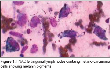
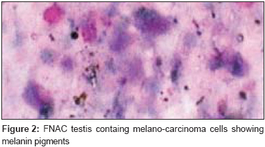
Journal of Cancer Research and Therapeutics, Vol. 4, No. 2, April-June, 2008, pp. 97-98 Case Report Primary melanoma of testis Katiyar RK, Singh Abhishek, Kumar Deepak Departrment of Radiotherapy, J.K. Cancer Institute, Kanpur Code Number: cr08027 Abstract Primary melanoma of testis is extremely rare and even the existence of such an entity is questioned. We present the case of a 60-year-old man with primary malignant melanoma in the testis. We report this case to emphasize the need for awareness of the possibility of the testis being the primary site in the patient with a melanoma and to underline the necessity of meticulous investigation of suspicious lesions of the testis in patients with or without a past history of malignant melanoma. Keywords: Melanoma, primary, testis Malignant melanoma, a malignant neoplasm that arises from melanocytes, is relatively rare and accounts for only 1-2% of all malignancies. However, the incidence is increasing rapidly, with a 300% increase being noted over the past 40 years. The incidence of the disorder varies in different populations and is about 10 times more common in white than in nonwhite populations. A primary cause of melanoma is believed to be sun exposure, and both UVA and UVB are suspected to be carcinogenic. At highest risk are people with fair complexions who have had intermittent sun exposure and a history of severe sunburns. Primary melanoma of testis is extremely rare and even the existence of the entity is questionable. We present the case of a 60-year-old man with primary malignant melanoma of the testis. We report this case to emphasize the need for awareness of the possibility of testis being the primary site in the melanoma patient and to underline the necessity of meticulous investigation of suspicious lesions of the testis in patients with or without a past history of malignant melanoma. Case Report A 60-year-old male presented with gradually increasing swelling in the left and right testis for 2 years and 6 months, respectively. Besides the testicular swellings, the patient also had a progressively increasing swelling in the left inguinal region for the past one year, which had burst open with blackish material coming out from it. This was followed by the appearance of a reddish colored spot, first on left shoulder and then gradually all over the body, which turned black with time. He also complained of fever off and on, anorexia, and weight loss for 4 months. Physical examination revealed an enlarged, 10 x 7 cm, left testis and a right testis sized 5 x 4.5 cm, both testes were firm to hard in consistency, with no changes in the overlying skin. There was a swelling in the left inguinal region measuring 7 x 7 cm in size that was variable in consistency. Another lymph node of about 5 x 5 cm size was also present in the same region, which was hard in consistency and nontender. The right inguinal region had a firm, mobile, and nontender lymph node of about 1 x 1.5 cm size. Black colored nodules, with size varying from 1-1.5 cm, were present over the trunk and chest; these nodules were raised above the skin surface, firm, and nontender. On abdominal examination, a nontender mild hepatomegaly was found. No other possible site for a primary tumor was identified. The hematological tests showed Hb: 7 gm%; WBC count: 4600/mm 3 ; DLC: P - 67, L - 25, E - 03, M - 05, B - 00; RBC count: 2.66 million/mm 3 ; and platelets: 4,85,000/mm 3 . Liver function tests were within normal limits. HBsAg was negative. Serum protein and electrolytes were within normal limits. The AFP level was 7 kU/l and the β-HCG level was 2 U/l. Ultrasonography (whole abdomen) revealed mild hepatomegaly with multiple sizeable solid (echogenic) as well as partially cystic focal liver lesions suggestive of secondaries; there was also mild splenomegaly. Multiple retroperitoneal lymph nodes were present. All the pelvic lymph nodes showed central hypoechogenicity suggestive of central necrosis. Bilateral hydronephrosis was present without any focal renal mass. No ascites was present. No abnormality was found in any other viscera. Proctoscopic examination found no abnormality. X-ray of the chest and the paranasal sinuses were normal. Color Doppler ultrasound of the scrotum revealed asymmetrical enlargement of both testes due to multiple, well-circumscribed, hypoechoic lesions with mild hydrocele. The lesions were echogenically heterogeneous and a few of them showed central necrosis. The epididymis was bulky bilaterally, with the right epididymis being more so than the left. There was evidence of a large lymph node mass lesion with heterogeneous echogenicity in the left inguinal region that was suggestive of lymph node secondaries. FNAC of the lesions in the liver, testis, and skin was done, which revealed malignant melanoma at all sites [Figure - 1] and [Figure - 2]. The patient′s general condition was poor. Surgery was not possible due to extensive disease. The disease followed an aggressive course and the patient died within a month while he was being managed conservatively. Discussion In 5-10% of cases, melanoma arises from sites other than the skin; these sites include the oral cavities, nasal sinuses, genitalia, and rectum. Melanoma may also occur in the uveal tract and the retina of the eye. However, no case of a primary in the testis has been reported till date. Theoretically, according to histogenesis, melanoblasts cannot be demonstrated in organs of mesodermal origin. Since the testis is an organ of mesodermal origin, it is unlikely that a melanoma can arise from this organ or from any of the other visceral structures. On the other hand, non-neoplastic melanoblasts resulting from migration of melanin-producing cells from the neural crest to mesodermal derivatives during embryologic development explains the presence of melanocytes in the testis and supports the possibility of a primary melanoma developing at this rare site. There have been a few cases reported of primary as well as metastatic melanoma in the gall bladder but no case of primary or metastatic melanoma in the testis has been reported till date.Copyright 2008 - Journal of Cancer Research and Therapeutics The following images related to this document are available:Photo images[cr08027f2.jpg] [cr08027f1.jpg] |
| |||||||||
