search
for
 About Bioline  All Journals  Testimonials  Membership  News


Journal of Cancer Research and Therapeutics
Medknow Publications on behalf of the Association of Radiation Oncologists of India (AROI)
ISSN: 0973-1482 EISSN: 1998-4138
Vol. 4, Num. 3, 2008, pp. 111-115

Journal of Cancer Research and Therapeutics, Vol. 4, No. 3, July-September, 2008, pp. 111-115

Original Article

Amelioration of cisplatin induced nephrotoxicity in Swiss albino mice by Rubia cordifolia extract

Department of Radiation Biology, Amala Cancer Research Centre, Amala Nagar, Thrissur-680 555, Kerala
Correspondence Address:Amala Cancer Research Centre, Trichur 680555, Kerala
ckknair@yahoo.com

Code Number: cr08034

Abstract

Background: Cisplatin is one of the most effective chemotherapeutics against a wide range of cancers including head, neck, ovarian and lung cancers. But its usefulness is limited by its toxicity to normal tissues, including cells of the kidney proximal tubule. The purpose of the present study is to investigate whether the hydro-alcoholic extract of Rubia cordifolia could decrease the intensity of toxicity in Swiss albino mice.
Materials and Methods:
Cisplatin at a dose of 12 mg/kg body wt was administered intraperitoneally to Swiss albino mice. Another set of animals was given hydro-alcoholic extract of Rubia cordifolia at different doses along with cisplatin treatment. The antioxidant levels, serum creatinine, serum urea etc. were analyzed.
Results: The extract could significantly decrease the cisplatin induced nephrotoxicity as inferred from the tissue antioxidant status in the drug administered animals. Remarkable change was observed in serum creatinine and urea levels. Lipid peroxidation in the kidney and liver tissues was also considerably reduced in Rubia cordifolia extract treated animals.
Conclusion: Hydro-alcoholic extracts of Rubia cordifolia are effective in reducing the renal damage caused by the cancer chemotherapeutic drug cisplatin. Since Rubia cordifolia has been in use as an important ingredient in the traditional Ayurvedic system of medicine, it could be safe and beneficial to use this herbal extract as an adjuvant to ameliorate renal damage in patients undergoing cancer chemotherapy with cisplatin.

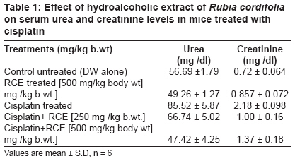
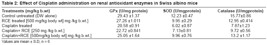
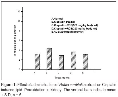
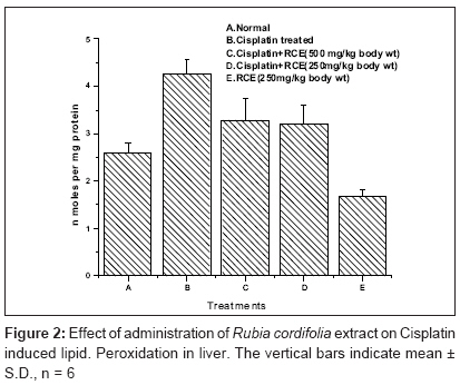
Keywords: Antioxidants, Cisplatin, nephrotoxiciity, Rubia cordifolia

Introduction

Cancer is one of the most dreaded diseases and currently taking the heaviest toll of human lives, with distant hope of finding an effective cure unless detected and treated in early stages. Chemotherapy and radiotherapy are the most common modalities of cancer treatment. Cisplatin (Cis-diammino dichloro platinum II) is currently one of the most important chemotherapeutic drugs used in treatment of a wide range of solid tumors - head, neck, ovarian and lung cancers. However, the clinical usefulness of this drug is limited due to nephrotoxicity induction, a side effect that may produced in various animal models. Cisplatin gets accumulated in the tubular epithelial cells of proximal kidney tubule, causing nephrotoxicity, characterized by morphological destruction of intra cellular organelles, cellular necrosis, loss of microvilli, alterations in the number and size of the lysosomes and mitochondrial vacuolization, followed by functional alterations including inhibition of protein synthesis, GSH depletion, lipid peroxidation and mitochondrial damage.

Several distinct mechanisms have been proposed for cisplatin cytotoxicity in renal tubule cells, including direct DNA damage, [1] activation of caspase [2] mitochondrial dysfunction, [3] formation of reactive oxygen species, [4] effects on the endoplasmic reticulum [5] and activation of TNF-a mediated apoptotic pathways. It has also been reported that cisplatin induced nephrotoxicity is closely associated with an increase in lipid peroxidation in the kidney. In addition, cisplatin has been found to lower the activities of antioxidant enzymes and to induce depletion of GSH.

A large number of studies have reported the beneficial effects of a variety of antioxidants in cisplatin induced nephrotoxicity. [6],[7] Agents such as SOD, dimethyl thiourea and GSH have been shown to reduce the degree of renal failure and tubular cell damage when administered simultaneously with cisplatin in rats. [4],[8],[9] Much attention has been given to the possible role of dietary antioxidants in protecting the kidney against cisplatin induced nephrotoxicity. There is a large body of evidence on the chemoprotecting activities of vitamin C, E, curcumin, selenium, bixin and other dietary components that scavenge free radicals induced by exposure to cisplatin. [10],[11],[12]

The potential of herbs and other plant-based formulations has increasingly been recognized in prevention and treatment of human diseases including cancer. The emerging integrative model of cancer treatment recognizes the importance of botanical medicine. The principles underlying herbal medicine are relatively simple, although they are quite distinct from conventional medicine. Here, efforts are made to exploit the nephroprotective effect of an ethnomedicinal plant, Rubia cordifolia .

Rubia cordifolia , otherwise known as Indian madder, belongs to the family Rubiaceae . [13] Roots contain resinous and extractive matter, gum, sugar, coloring matter and salts of lime. Coloring matter consists of a red crystalline principle - purpurin, a yellow principle glucoside - manjistin, besides garancin, alizarin (orange-red) and xanthine (yellow). Anthraquinones, pentacyclic triterpenes, quinines, and cyclic hexapeptides and diethyl esters are also reported. [14],[15]

Materials and Methods

Chemicals
Nitroblue tetrazolium (NBT), riboflavin, reduced glutathione (GSH), 5-5′ dithiobis 2-nitro benzoic acid (DTNB) were obtained from Sisco Research Laboratories Pvt Ltd, Mumbai, India and Cisplatin from Samarth Pharma Pvt Ltd, Mumbai. Thiobarbituric acid (TBA) was purchased from Hi-media Laboratories, Mumbai, India. All other chemicals and reagents used in this study were of analytical grade.

Animals
Inbred 4-6 week-old male Swiss Albino mice (20-25 gm) were obtained from Sri Venketeswara Enterprises, Bangalore. They were kept in well-ventilated cages under standard conditions of temperature, pressure and humidity. The animals were provided with normal mouse chow (Sai Durga Feeds and Foods, Bangalore) and water ad libitum. All animal experiments conducted during the present study got prior permission and followed the guidelines of Institutional Animal Ethics Committee (IAEC).

Preparation of hydro-alcoholic extract of Rubia cordifolia
Authenticated dried roots of Rubia cordifolia were purchased from Amala Ayurvedic Hospital and Research Centre and powdered. The powder was extracted with 70% ethanol at room temperature. Extract was filtered through Whatmann No. 1 filter paper and the supernatant was evaporated using rotary evaporator at 45 O C and the final liquid suspension was lyophilized to get a powder with 11% yield, hereafter referred as RCE ( Rubia cordifolia extract). The powder was dissolved in distilled water at desired concentrations and administered orally by gavage to animals.

Determination of nephroprotection by Rubia cordifolia
Animals were divided into five groups of six animals each. Group I treated with vehicle (distilled water) was kept as normal. Group II injected with a single dose of cisplatin (12 mg/kg body weight; i.p.) was kept as control. Group III was administered with Rubia cordifolia extract (RCE) 250 and 500 mg/kg body weight along with cisplatin treatment. Group IV was given RCE at a dose of 500 mg/kg body wt alone. The extract was administered by oral gavage 1 h before and at 24 h and 48 h after cisplatin injection. Seventy-two hours after the cisplatin injection, animals were sacrificed using ether- anesthesia; blood samples were collected by heart puncture for measuring serum urea and serum creatinine levels. Kidneys were quickly removed and washed with ice-cold normal saline and homogenates (10% w/v) were prepared in PBS. A part of the homogenate was used for the estimation of reduced glutathione (GSH) and lipid peroxidation. The remaining homogenate was centrifuged at 5000 xg for 10 minutes at 4 o c, after removal of the cell debris, supernatant was used for the assay of Super oxide dismutase (SOD), Catalase (CAT), Glutathione Peroxidase (GPx) etc.

Serum Creatinine is determined by alkaline picric acid method using a diagnostic kit (Agappe Diagnostic Pvt Ltd; Ernakulam, Kerala, India). Serum urea was determined by diacetylmonoxime (DAM) reagent (modified Berthelot methodology) using a diagnostic kit (Agappe Diagnostic Pvt Ltd; Ernakulam, India).

Determination of tissue reduced glutathione (GSH)
It is the most abundant intracellular thiol compound present in virtually all mammalian cells. GSH participates in a reaction that destroys H 2 O 2 , organic peroxides, free radicals and certain foreign compounds. GSH reacts with DTNB and gets reduced to a yellow colored complex which has an absorption maximum at 412 nm. [16]

Determination of tissue glutathione peroxidase (GPx) activity
Glutathione peroxidase is the general name of an enzyme family with peroxidase activity, whose main role is to protect the organism from oxidative damage. The biochemical function of GPx is to reduce lipid hydro peroxides to their corresponding alcohols and to reduce free H 2 O 2 to H 2 O. GPx remaining is measured using DTNB, which gives a yellow colored complex. [17]

Determination of tissue superoxide dismutase activity
SOD is a metalloenzyme with its active center occupied by copper or zinc, sometimes manganese (or) iron. SOD plays an important role in protection of all aerobic life systems, against oxygen toxicity. SOD catalyzes the dismutation of superoxide into O 2 and H 2 O 2 . The assay is based on the ability of the enzyme to inhibit the reduction of nitro blue tetrazolium (NBT) by superoxide, which is generated by the reaction of photo reduced riboflavin with O 2 . [18]

Determination of in vivo Lipid Peroxidation (LPO)
Malondialdehyde (MDA) is a secondary product of lipid peroxidation and is used as an indicator of tissue damage. The MDA reacts with TBA reagent to from a pink colored product which has absorption maxima at 532 nm. The concentrations of malondialdehyde (MDA) as indices of lipid peroxidation were assessed according to the method of Buege and Aust. [19]

Determination of catalase activity
Catalase activity was determined from the rate of decomposition of H 2 O 2, monitored by decrease of 240 nm following the addition of tissue homogenate. [20]

Determination of tissue protein
The protein estimation was done using Lowry′s method with BSA as standard. [21]

Results

The data presented in [Table - 1] reveals that the values of serum urea and creatinine were significantly elevated in the cisplatin treated group. Cisplatin treatment resulted in a two-fold increase in the values of serum urea and creatinine levels as compared to that of the untreated control group. The administration of RCE to cisplatin treated mice could restore the elevated levels of urea and creatinine to that of the untreated control levels.

The major antioxidant enzymes such as GPx, SOD and Catalase were found to be decreased in cisplatin treated animals and per oral administration of RCE could elevate these levels as can be realized from the data presented in [Table - 2].

From [Figure - 1] and [Figure - 2] it can be seen cisplatin treatment resulted in increased peroxidation of lipids in the kidney and liver - tissues of mice and administration of RCE resulted in inhibition of cisplatin-induced peroxidation of lipids in these tissues. The protection against cisplatin induced toxicity could stem from the potent antioxidant activity of RCE. Inhibition of LPO in biomembranes has been caused by antioxidants present in the plant extract. From the above results, it can be inferred that the extract of Rubia cordifolia offers protection against cisplatin induced oxidative stress in renal tissues.

Discussion

Nephrotoxicity is one of the major side effects of cisplatin. Although several studies have been performed to elucidate the molecular mechanisms that cause cisplatin nephrotoxicity, the factors responsible for this are not fully understood. Recently, induction of oxidative free radicals has been implicated in this process. [22] Different strategies have been proposed to inhibit cisplatin induced toxicity. The development of therapies designed to prevent the damaging actions of free radicals may influence the progression of oxidative renal damage induced by cisplatin.

The major antioxidant enzymes such as GPx, SOD and Catalase were found to be decreased in cisplatin treated animals and per oral administration of RCE could elevate these levels [Table - 2]. ROS such as hydrogen peroxide, the superoxide anion, and hydroxyl radicals are generated under normal cellular conditions and are immediately detoxified by endogenous antioxidants, like GSH, catalase and superoxide dismutase, but excessive ROS accumulation by cisplatin causes an antioxidant status imbalance and leads to lipid peroxidation and GSH depletion. [23]

The basic effect of cisplatin induced toxicity on the cellular membrane is believed to be peroxidation of membrane lipids. The depletion of glutathione at early intervals in treated animals may be due to its utilization in large amounts to combat the acute cisplatin induced free radical damage, as glutathione is a major nonenzymatic antioxidant. The measurement of lipid peroxidation as thiobarbituric acid reacting substances (TBARS) is a convenient method to monitor oxidative damage in tissues. Reactive oxygen species cause peroxidation of membrane lipids with devastating effect on functional states. The preservation of cellular membrane integrity depends on protection or repair mechanisms capable of neutralizing oxidative reactions. Our data show that cisplatin induced malondialdehyde (MDA) production was significantly decreased by the p.o. administration of RCE in vivo and it also attenuated cisplatin induced GSH depletion in mice. It has been suggested that cisplatin is able to generate ROS and that it inhibits the activities of antioxidant enzymes in renal tissues. [24] In the present study the reduced activities of GPx, SOD and Catalase in kidneys of mice treated with cisplatin were restored by administration of RCE to a considerable extent indicating the ability of RCE to eliminate oxidative stress.

Cisplatin has been thought to bind to the renal base transport system. Cisplatin induces hypomagnesemia through its renal toxicity possibly by a direct injury to mechanisms of magnesium reabsorption in the ascending limb of the loop of Henle as well as the distal tubule. Cisplatin preferentially accumulates in cells of the S3 segment of the renal proximal tubules and is toxified to form a reactive metabolite intracellularly by hydration. The primary symptoms of cisplatin nephrotoxicity are inhibition of protein synthesis and intracellular GSH and protein-SH depletion, resulting in lipid peroxidation and mitochondrial damage. [23] The peroxidation of membrane lipids may account for its nephrotoxicity. [25] Available evidence suggests that cisplatin exerts its nephrotoxic effects by the generation of free radicals. [26],[27],[28] GSH and protein-SH form the major cellular anti-oxidant defense systems, which control lipid peroxidation. From these pathomechanisms of cisplatin nephrotoxicity, it is clear that the nephrotoxicity of cisplatin involves reactive radicals. Thus the reasonable cellular-protective agents against cisplatin toxicity may have at least some antioxidant properties to prevent GSH depletion and/or scavenge the intracellular reactive oxygen species.

The present observations support the hypothesis that the mechanism of nephrotoxicity is related to the depletion of the antioxidant defense system. Cisplatin treatment has been shown to induce loss of copper and zinc in the kidneys. The decrease in SOD activity in renal tissues following cisplatin administration might be due to the loss of copper and zinc.[29] The activity of Catalase and GPx is also found to decrease after cisplatin administration resulting in the decreased ability of the kidney to scavenge toxic hydrogen peroxide and lipid peroxides. The results from the present study indicate that the extract (RCE) significantly reduced the depletion of GSH levels and antioxidant enzyme activity in the renal cortex of mice treated with cisplatin.

Numerous studies have shown that cisplatin induces renal damage by free radical generation. Hence antioxidants and free radical scavengers of natural and synthetic origin might provide nephroprotection in cisplatin induced renal injury. [30] It has been reported that anthraquinones are the major components present in Rubia cordifolia. The genus Rubia is a rich source of anthraquinones. For example, many anthraquinones such as 1-hydroxy-2-methylanthraquinone and nordamnacanthal had been isolated from the roots of Rubia cordifolia. [31] The antioxidant properties of anthraquinones (AQs) and anthrones were evaluated using different model systems. Anthraquinones possess good antioxidant activity due to its reducing power and scavenging effects on hydroxyl radicals.

It has been reported that Rubia cordifolia extract possesses potent free radical scavenging property. [31] The present study demonstrates the potent antioxidant properties of the extract. Hence, it may be concluded that the mechanism of nephroprotection by Rubia cordifolia extract in cisplatin treated mice could be due to the antioxidant and free radical scavenging activity.

References

1.Leibbrandt ME, Wolfgang GH, Metz AL, Ozobia AA, Haskins JR. Critical subcellular targets of cisplatin and related platinum analogs in rat renal proximal tubule cells. Kidney Int 1995;48:761-70  Back to cited text no. 1  [PUBMED]  
2.Kaushal GP, Kaushal V, Hong X, Shah SV. Role and regulation of activation of caspases in cisplatin induced injury to renal tubular epithelial cells. Kidney Int 2001;60:1726-36.  Back to cited text no. 2  [PUBMED]  [FULLTEXT]
3.Sugiyama S, Hayakawa M, Kato T, Hanaki Y, Shimizu K, Ozawa T. Adverse effects of anti-tumor drug, cisplatin on rat kidney mitochondria: Disturbances in glutathione peroxidase activity. Biochem Biophys Res Commun 1989;159:1121-7  Back to cited text no. 3  [PUBMED]  [FULLTEXT]
4.Matsushima H, Yonemura K, Ohishi K, Hishida A. The role of oxygen free radicals in cisplatin induced acute renal failure in rats. J Lab Clin Med 1998;131:518-26.  Back to cited text no. 4  [PUBMED]  [FULLTEXT]
5.Baliga, R., Liu, H. Activation of caspase 12 by cisplatin (CP) induced endoplasmic reticulam (ER) stress mediates apoptosis in LLC-PK1 cells. Am Soc Nephrol 2004;15:39.  Back to cited text no. 5    
6.Baek SH, Piao XL, Lee UJ, Kim HY, Park JH. Reduction of cisplatin-induced nephrotoxicity by Ginsenosides isolated from processed Ginseng in cultured renal tubular cells. Biol Pharm Bull 2006;29:2051-5.  Back to cited text no. 6  [PUBMED]  [FULLTEXT]
7.Atessahin A, Yilmaz S, Karahan I, Ceribasi AO, Karaoglu A. Effects of lycopene against cisplatin-induced nephrotoxicity and oxidative stress in rats. Toxicology 2005;212:116-23.  Back to cited text no. 7  [PUBMED]  [FULLTEXT]
8.Sadzuka Y, Shoji T, Takino Y. Effect of cisplatin on the activities of enzymes which protect against lipid peroxidation. Biochem Pharmacol 1992;43:1872-5.  Back to cited text no. 8  [PUBMED]  [FULLTEXT]
9.McGinness JE, Proctor PH, Demopoulos HB, Hokanson JA, Kirkpatrick DS. Amelioration of cisplatin nephrotoxicity by orgotein (SOD). Physiol Chem Phys 1978;10:267-77.  Back to cited text no. 9  [PUBMED]  
10.Antunes LM, Darin JD, Bianchi MD. Protective effects of vitamin C against cisplatin induced nephrotoxicity and lipid peroxidation in adult rats, a dose dependent study. Pharmacol Res 2000;41:405-11  Back to cited text no. 10  [PUBMED]  [FULLTEXT]
11.Antunes LM, Darin JD, Bianchi Nde L. Effects of the antioxidants curcumin or selenium on cisplatin induced nephrotoxicity and lipid peroxidation in rats. Pharmacol Res 2001;43:145-50.  Back to cited text no. 11  [PUBMED]  [FULLTEXT]
12.Silva CR, Greggi Antunes LM, Bianchi ML. Antioxidant action of bixin against cisplatin induced chromosome aberrations and lipid peroxidation in rats. Pharmacol Res 2001;86:271-8.  Back to cited text no. 12    
13.Tripati YB, Singh AV Role of Rubia cordifolia Linn. in radiation protection. Indian J Exp Biol 2007;45:620-5  Back to cited text no. 13    
14.Singh R Geetanjali. Isolation and synthesis of anthraquinones and related compounds of Rubia cordifolia . J Serb Chem Soc 2005;70:937-42.  Back to cited text no. 14    
15.Joharapurkar AA, Zambad SP, Wanjari MM, Umathe SN. In vivo evaluation of antioxidant activity of alcoholic extract of Rubia cordifolia linn. and its influence on ethanol-induced Immunosuppression. Indian J Pharmacol 2003;35:232-6  Back to cited text no. 15    
16.Moron MA, Depicrre JW, Mannervick B. Levels of glutathion, glutathione-s -transferase activities in rat liver. Biochem Biophys Acta 1979;582:67-8  Back to cited text no. 16    
17.Hafeman DG, Sunde RA, Hoekstra WG. Effect of dietary selenium on erythrocyte and liver glutathione peroxidase in the rat. J Nutr 1974;104:580-7  Back to cited text no. 17  [PUBMED]  [FULLTEXT]
18.McCord JM, Fridovich I. Superoxide dismutase enzyme function for erythrocuprein (hemocuprein). J Biol Chem 1969;244:6049-55.  Back to cited text no. 18    
19.Buege JA, Aust SD. Microsomal lipid peroxidation. Met Enzymol 1978;52:302-10.  Back to cited text no. 19    
20.Aebi H. Catalase. In: Bergmeyer HU, editor. Methods in enzymatic analysis, Vol. 3. New York: Academic Press; 1983. p. 276-286  Back to cited text no. 20    
21.Lowry OH, Rosebrough NJ, Farr AL, Randall RJ. Protein measurement with folin phenol reagent. J Biol Chem 1951;193:265-75.  Back to cited text no. 21  [PUBMED]  [FULLTEXT]
22.Baldew GS, McVie JG, van der Valk MA, Los G, de Goeij JJ, Vermeulen NP. Selective reduction of cis-diamminedichloroplatinum(II) nephrotoxicity by ebselen. Cancer Res 1990;50:7031-6.  Back to cited text no. 22  [PUBMED]  [FULLTEXT]
23.Kim YH, Kim YW, Oh YJ, Back NI, Chung SA, Chung HG, et al . Protective effect of the ethanolic extract of the roots of Brassica rapa on cisplatin induce nephrotoxicity in LLC-PK1 cells and rats. Biol Pharm Bull 2006;29:2436-41  Back to cited text no. 23  [PUBMED]  [FULLTEXT]
24.Naziroglu M, Karaogl A, Aksoy AO. Selenium and high dose vitamin E administration protects cisplatin induced oxidative damage to renal, liver and lens tissues in rats. Toxicology 2004;195:221-30  Back to cited text no. 24    
25.Safirstein R, Miller P, Guttenplan JB. Uptake and metabolism of cisplatin by rat kidney. Kidney Int 1984;25:753-8.  Back to cited text no. 25  [PUBMED]  
26.Trakshel GM, Maines MD. Characterization of glutathione S-transferases in rat kidney: Alteration of composition by cis-platinum. Biochem J 1988;252:127-36.  Back to cited text no. 26  [PUBMED]  [FULLTEXT]
27.Ishikawa M, Takayanagi Y, Sasaki K. Enhancement of cisplatin toxicity by buthionine sulfoximine, a glutathione depleting agent in mice. Res Commun Chem Pathol Pharmacol 1990;67:131-41.  Back to cited text no. 27  [PUBMED]  
28.Uslu R, Bonavida B. Involvment of the mitochondrion respiratory chain in the synergy achieved by treatment of human ovarian carcinoma cell lines with both tumor necrosis factor-alpha and cis-diamminedichloroplatinum. Cancer 1996;77:725-32.  Back to cited text no. 28  [PUBMED]  
29.Sharma RP. Interactions of cisplatin with cellular zinc and copper in liver and kidney tissues. Pharmacol Res Commun 1985;17:197-206.  Back to cited text no. 29  [PUBMED]  
30.Maliakel DM, Kagiya TV, Nair CK. Prevention of cisplatin-induced nephrotoxicity by glucosides of ascorbic acid and alpha-tocopherol. Exp Toxicol Pathol 2008 (in Press).   Back to cited text no. 30    
31.Tessier AM, Delaveau P, Champion B. New anthraquinones in Rubia cordifolia root. Planta Med 1981;41:337-43.  Back to cited text no. 31  [PUBMED]  

Copyright 2008 - Journal of Cancer Research and Therapeutics


The following images related to this document are available:

Photo images

[cr08034t1.jpg] [cr08034t2.jpg] [cr08034f2.jpg] [cr08034f1.jpg]
Home Faq Resources Email Bioline
© Bioline International, 1989 - 2024, Site last up-dated on 01-Sep-2022.
Site created and maintained by the Reference Center on Environmental Information, CRIA, Brazil
System hosted by the Google Cloud Platform, GCP, Brazil