 |
| About Bioline | All Journals | Testimonials | Membership | News |
|
||||||
|
||||||
Journal of Cancer Research and Therapeutics, Vol. 6, No. 1, January-March, 2010, pp. 3-4 Invited Editorial Evolution of radiation oncology: Sharp gun, but a blurred target Anusheel Munshi, Jai Prakash Agarwal Department of Radiation Oncology, Tata Memorial Hospital, Mumbai, India Code Number: cr10002 DOI: 10.4103/0973-1482.63550 "In the long history of humankind (and animal kind, too) those who learned to collaborate and improvise most effectively have prevailed." …….…"It is not the strongest of the species that survives, nor the most intelligent that survives. It is the one that is the most adaptable to evolving change"…. Charles Darwin . As per the legendary law of Charles Darwin, natural life, depending on the need of the species concerned, evolves in a stepwise and orderly fashion. [1] By and large, medicine is no exception to this general rule, with one milestone being the leading light for the next and higher frontier. Over the past two decades, Radiation Oncology too has taken huge technology strides. The traditional low energy orthovoltage machines have become a curiosity. Cobalt machines, which replaced the orthovoltage machines have bowed down to elegant and sophisticated linear accelerators (LA). Of late, LAs themselves have undergone radical (almost biannual!) transformation from machines capable of delivering simple square or rectangular 4 MV/6 MV beams to the current C arm LA which boasts of multileaf collimators, arc treatment, asymmetric jaws and dynamic motion capabilities. Taking a cue from computerized tomography (CT) scanners, helical rotational machines have mushroomed across the globe. [2] Intensity Modulated Radiotherapy and Image Guided Radiotherapy are the latest feathers in the cap for radiation delivery. These technologies allow us to create amazing patterns of dose fluence across the target area, allowing to deliberately inhomogenise the dose intensity depending on the risk of disease recurrence for a sub volume within the target area. In sum, modern radiation oncology set up is easily capable of precision delivery to a sub millimeter level. [3] However, from an unbiased and neutral perspective, this rapid development of technology is intriguing. [4] This puzzle can be understood by revising the fundamentals of clinical practice in radiation oncology. In radiation oncology parlance, the gross tumor volume (GTV) denotes the gross visible or palpable tumor while the clinical target volume (CTV) determines the area of microscopic disease around the gross tumor. Finally, the planning target volume (PTV) indicates the extra margin to account for external set up and internal motion of the organs. Traditional radiotherapy essentially consisted of parallel pair or simple field arrangements with generous margins based on surface anatomy or fluoroscopic visualization. These liberal portals included both the disease site (GTV, CTV, PTV) while often including many areas of organs at risk (OAR) as well. True, in many instances this leads to considerable side effects and limited delivery of higher doses. Ostensibly, as a step forward, the present day radiotherapy planning involves CT slice based contouring of the tumor (GTV, CTV and PTV) and the OAR. [5] Efforts are being made to account for tumor motion and respiratory movement and many centers are already treating patients with elegant techniques of respiratory gating, breath control and so on. However, in a rational order of evolution and development, precision delivery should have followed or paralleled the evolution of contouring. While newer imaging modalities in radiology have improved GTV imaging, serious issues regarding contouring of CTV and PTV remain. [6] In patients with an intact GTV a "suitable margin" is given by the radiation oncologist to account for microscopic spread of disease (or the CTV). Literature for the extent of this margin (derived from pathological series, imaging data, wisdom earned from pattern of recurrences) is limited for most sites. A patient who lands up after neoadjuvant chemotherapy or has been operated upon (thus distorting the muscle and the fat planes) is a perfect recipe for further and added uncertainty; especially after chemotherapy, the perineal dilemma is between hypothetically drawing the initial tumor from pre chemotherapy scans versus taking the post chemotherapy volume the target. Finally, the rules for giving the PTV margin (to account for internal motion and daily set up variations) are far from being ironed out. The proof of pudding for the aforesaid dilemma is the large interobserver variation seen for various studies involving contouring in recent studies. [7] Put simply, this leaves the radiation oncologist with a super sharp gun but a highly blurred target [Figure - 1]. What follow is a peculiar situation which the radiation oncology community should be cognizant of - being less forgiving in view of precision delivery, modern radiotherapy is at a higher risk of erroneously (and precisely!) sparing the tumor or conversely irradiating the OARs in some cases. [8],[9] This is reflected in warning bells sounded by reports of periparotid recurrences (in an attempt to give tighter margins aiming to save the parotid) in head and neck cancer patients treated with intensity modulated radiotherapy. [10] Similar reports could soon emerge for other sites. Historically, radiology and radiation oncology started as a single department and parted ways down the line. More and more of radiotherapy planning is, however, currently being done based on CT slices. Often these images are fused with magnetic resonance imaging (MRI), Positron emission tomography (PET) or perfusion images. It may be the perfect time for a partial reunion of sorts. In other words, time has come to stop working on perfecting the guns (at least for the time being!) and concentrate efforts on figuring out the target. Till then, the ghost of Darwin would continue to haunt us. References
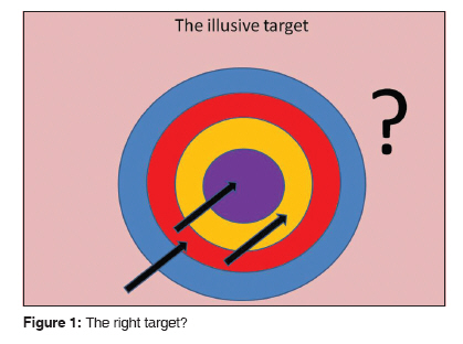
Copyright 2010 - Journal of Cancer Research and Therapeutics The following images related to this document are available:Photo images[cr10002f1.jpg] |
| |||||||||
