 |
| About Bioline | All Journals | Testimonials | Membership | News |
|
||||||
|
||||||
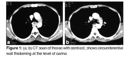
Journal of Cancer Research and Therapeutics, Vol. 6, No. 1, January-March, 2010, pp. 100-101 Case Report Spindle cell sarcoma of esophagus: A rare case presentation Lokesh V., Naveen T., Y. S. Pawar Department of Radiation Oncology, Kidwai Memorial Institute of Oncology, Dr MH Marigowda Road, Hosur Road, Bangalore-560 029, India Code Number: cr10023 DOI: 10.4103/0973-1482.63558 Abstract Sarcomas of the esophagus, including carcinosarcoma, are rare neoplasm cases and comprise 0.1-1.5% of all esophageal tumors. Leiomyosarcoma is the most common of the pure mesenchymal tumors of the esophagus, but sarcomas with combined histological types such as carcinosarcoma occur more frequently than pure sarcomas. We report a rare case of spindle cell sarcoma of esophagus in a 55-year-old woman, managed with radical radiotherapy alone.Keywords: Carcinosarcoma, esophagus, radiotherapy, sarcoma Introduction Sarcomas of the esophagus, including carcinosarcoma, are rare neoplasm cases and comprise 0.1-1.5% of all esophageal tumors. [1] Leiomyosarcoma is the most common of the pure mesenchymal tumors of the esophagus, but sarcomas with combined histological types such as carcinosarcoma occur more frequently than pure sarcomas. We report a rare case of spindle cell sarcoma of esophagus in a 55-year-old woman managed with radical radiotherapy alone. Case Report A 55-year-old woman presented with two months duration of dysphagia for solid food. She had no significant past medical history. Clinically, the patient was normal with KPS 60. Hemogram, biochemistry, chest X-ray (CXR) and ultrasonography (USG) abdomen were normal. Computed tomography (CT) scan was suggestive of ill-defined enhancing soft tissue density mass lesion from the level of post cricoid region up to the level of carina measuring approximately 7.2 cm, associated with skip lesions from the level of right pulmonary artery down up to 3.2 cm. Endoscopic examination of esophagus showed an ulcerative lesion on posterior wall of esophagus extending from 20 cm up to 25 cm from incisors. Histopathological examination of tissue showed spindle cells with more than five mitoses per highpower fields (HPF). Immunohistochemistry studies showed weak desmin positive, cytokeratin negative suggesting consistent features of malignant stromal tumor of smooth muscle origin. Considering the aggressive nature of the disease, guarded prognosis, limited outcome, general condition and radio resistant nature of the disease, patient was treated with radical radiotherapy to the total equivalent dose of 66 Gy (300 c Gy per fraction for two weeks followed by 200 c Gy per fraction for three weeks) The patient tolerated the treatment well with no normal tissue complications; three months post- radiotherapy, there was complete response of the tumor. Patient remained disease-free for 24 months and expired due to myocardial infarction. [Figure - 1]a and b Discussion Esophageal sarcomas are rare and occur most commonly as polypoid intraluminal masses. They may be divided into tumors with mixed epithelial and spindle cell characteristics such as carcinosarcoma and pure sarcomas of mesenchymal origin. Sarcomas of the esophagus, including carcinosarcoma, comprise 0.1-1.5% of all esophageal tumors. Leiomyosarcoma is the most common of the pure mesenchymal tumors of the esophagus, but sarcomas with combined histological types such as carcinosarcoma occur more frequently than pure sarcomas such as leiomyosarcoma. Carcinosarcoma, first named by Virchow in 1865, is more common and consists of intermingled malignant epithelial and sarcomatous components, both of which are known to metastasize. The application of antiepithelial markers (cytokeratin) would be of help to verify the absence/presence of epithelial component in this tumor with sarcoma/carcinosarcoma. [2] Like squamous cell carcinoma of the esophagus, carcinosarcoma occurs most commonly in middle-aged and elderly males with a history of smoking and/ or alcohol use. [3] Pure sarcomas of the esophagus are very rare. The most common of these is leiomyosarcoma. The diagnosis of a malignant soft tissue tumor (sarcoma), especially in gastrointestinal (GI) tract, gets riddled with a number of questions regarding the true nature of such a tumor. A large body of recent literature has addressed these questions and strongly recommended the application of immunocytochemistry, for the precise definition of sarcomas in the GI tract. [4],[5],[6],[7] Recent studies have shown that the presence of desmin and smooth muscle actin (SMA) is useful in confirming a histologic impression of entrapment of smooth muscle bundles. [4],[5],[6] Leiomyosarcoma usually occurs in the middle or distal portion of the esophagus. Esophageal leiomyosarcoma had, in the past, been classified as polypoid in 60% of cases and infiltrative in 40%. [8],[9] As for the radiographic findings in leiomyosarcoma of the esophagus, Levine et al. report that esophageal leiomyosarcoma has radiographic features similar to those of leiomyosarcoma found elsewhere in the GI tract. [10] It is important to be aware of the limitations of endoscopy in diagnosing esophageal leiomyosarcoma. Endoscopic biopsy specimens can be helpful if the mucosa overlying the tumor is ulcerated. If the overlying mucosa is intact, however, a superficial biopsy may yield a false-negative result. The first attempt of biopsy was unsuccessful in our patient too. The superficial nature of the biopsy specimens precludes an accurate histological diagnosis in many patients with this tumor. Leiomyosarcoma has a slow and indolent clinical course, followed by late recurrence, and eventual death of patients from the disease. Hematogenous metastasis was the cause of most of the cases of tumor recurrence and death. Conclusion In inoperable cases of esophageal sarcomas, radiotherapy may play an important role as primary modality of treatment. References
Copyright 2010 - Journal of Cancer Research and Therapeutics The following images related to this document are available:Photo images[cr10023f1.jpg] |
| |||||||||
