 |
| About Bioline | All Journals | Testimonials | Membership | News |
|
||||||
|
||||||
Journal of Cancer Research and Therapeutics, Vol. 7, No. 3, July-September, 2011, pp. 314-324 Original Article In vitro and in vivo targeted delivery of photosensitizers to the tumor cells for enhanced photodynamic effects Seema Gupta1, Bilikere S Dwarakanath2, NK Chaudhury2, Anil K Mishra3, K Muralidhar4, Viney Jain5 1 Division of Radiation Biosciences, Institute of Nuclear Medicine and Allied Sciences, Brig S K Mazumdar Road, Delhi, India; Department of Radiation Oncology, and Sylvester Comprehensive Cancer Center, University of Miami, Miami, FL 33136, USA, PMID: 22044814 DOI: 10.4103/0973-1482.87035 Background: Efficacy of photodynamic therapy can be enhanced by improving uptake, localization, and sub-cellular localization of sensitizers at the sensitive targets.
Keywords: 99m Tc labeling, biodistribution, liposome encapsulation, localization, monoclonal antibody, photodynamic treatment, photosensitizer, sub-cellular localization, targeted delivery, uptake
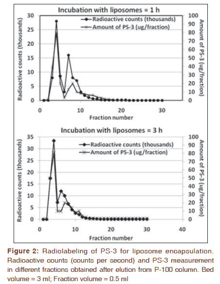
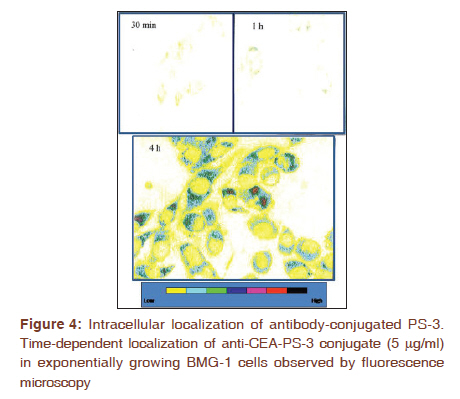
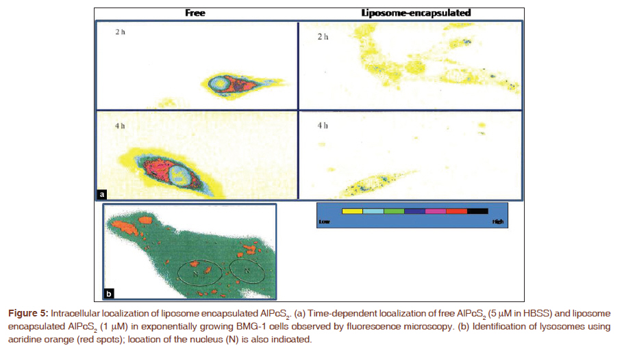
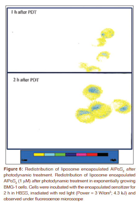
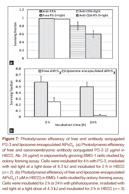
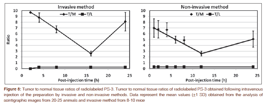
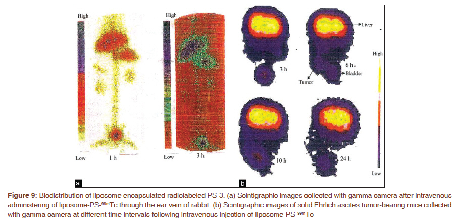
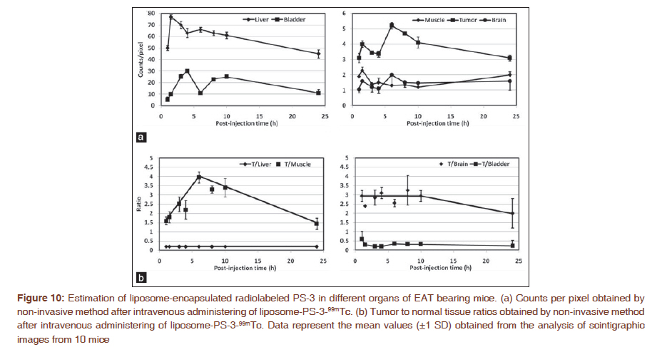
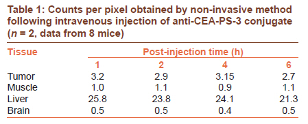
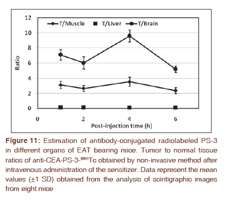
Introduction Efficacy of photodynamic treatment (PDT) depends on several factors such as uptake and localization of the photosensitizer, mode of light delivery, physiological status of the cells or the target tissue, and photophysical properties of the sensitizer like singlet and triplet quantum yields and life-times. Methods/techniques that help in increased accumulation and retention of the sensitizer in the target tissue could be one of the approaches to differentially enhance the damage to the tumor tissue as compared to the normal tissue. Also, the sub-cellular localization of the sensitizers at sites that are most sensitive to the photodynamic treatment is another important factor influencing the photodynamic efficacy, since singlet oxygen has a short half-life, and therefore can cause damage at or near the site of its production. Therefore, there is considerable amount of interest in investigations pertaining to the optimization of uptake, distribution, and localization of the sensitizers both in vitro and in vivo. Selective damage to the tumor can be achieved by increased accumulation and retention of the sensitizer in the tumor, [1],[2],[3],[4] development of better sensitizers, [5],[6] light sources and delivery systems, [7] and modulation of metabolism to selectively enhance the manifestation of PDT-induced damage in the tumor. [8],[9],[10],[11] New photosensitizers that absorb significantly in the far red region with higher yields of singlet oxygen and reduced localization in skin have been developed. [12],[13] Several delivery systems such as photosensitizer conjugates, dendrimers, micelles, liposomes, and nanoparticles, besides the application of photochemical internalization (PCI) technology for intracellular delivery of macromolecules have been recently described. [14],[15],[16] In the present study, localization, distribution, and targeting of the sensitizers, photosan-3 (PS-3) and disulfonated phthalocyanine (AlPcS 2 ) were modulated by encapsulating in liposomes and conjugating with monoclonal antibodies. Both liposome encapsulation and antibody conjugation resulted in localization of sensitizers at sensitive targets and reduced accumulation in the normal tissues, suggesting that both these delivery systems have potential to selectively enhance local tumor control following photodynamic treatment. Materials and Methods Chemicals The commercially available form of the photosensitizer, hematoporphyrin derivative, was obtained as PS-3 from SeeLab (Hamburg, Germany). Antibody to carcinoembryonic-antigen (CEA) was provided by CIM, Hawana, Cuba. This was conjugated to PS-3 using water-soluble activating agents, N-(3-dimethylaminopropyl)-N′-ethylcarbodiimide hydrochloride (EDC-C8H17N3·HCl) and N-Hydroxy sulfosuccinimide sodium salt (S-NHS-C4H4NNaO6S). Sephadex G-50 (Pharmacia) was used to separate conjugated PS-3 from free PS-3. Fluorescein Isothiocyanate (FITC)-conjugated secondary antibody (mouse anti-IgG) was from Sigma Chemical Co., USA, and stannous chloride (SnCl 2 , used as a reducing agent) from Glaxo Laboratories. Disulfonated aluminum phthalocyanine (AlPcS 2 ) was prepared and characterized in Institute of Nuclear Medical and Allied Science (INMAS), Delhi. [17] 99m Technetium for radioactive labeling was generated at INMAS, Delhi. DL-alpha-PC dipalmitoyl (C16:0) (1,2-dihexadecanyl-rac-glycero-3-phosphocholine, C 40 H 80 NO 8 P) (DPPC, SIGMA), cholesterol (5-cholesten-3beta-ol, C 27 H 46 O, SIGMA), dialysis tubing (cellulose membrane, SIGMA, Size- mol. wt. cut off 12000 or more), P-100 (Biogel), Osmium tetraoxide (Agar Scientific Ltd., UK) were used for liposome preparation and characterization. All other chemicals were of analytical grade and obtained from Glaxo Laboratories (Qualigens) or E-Merck, India. Tumor cell line Murine Ehrlich ascites tumor (EAT, F15) cells were obtained from University of Frankfurt, Germany [18] and used for animal studies. Human cerebral glioma cell line (BMG-1; DNA index = 0.95; wild-type p53), established from a mixed glioma [19] was used for in vitro studies. Monolayer BMG-1 cells were grown in Dulbecco′s modified Eagle′s medium (DMEM) with 5% fetal calf serum (FCS), penicillin (100 units/ml), streptomycin (50 mg/ml) and nystatin (2 mg/ml). Stock cultures were sub-cultured every third day after harvesting the cells with 0.05% trypsin and seeding 8 × 10 3 cells/cm 2 in tissue culture flasks to maintain the cells in the expo-nential phase. All experiments were carried out with exponentially growing cells. Animal models Swiss albino, strain ′A′ male mice inbred at INMAS were used as experimental animal model. EAT cells were grown in the peritoneal cavity of three-month-old male ′A′ strain mice and harvested on every seventh day after inoculation. Viable cells excluding 0.5% trypan blue were counted using Neubauer chamber and 25 million cells were injected intraperitoneally for propagation. For implantation of solid tumors, EAT cells (15 × 10 6 ) were injected subcutaneously in 0.1-0.3 ml volume in the right hind leg of the mice. After tumor implantation, animals were observed for tumor growth. Tumor diameters were measured in three mutually perpendicular directions with the help of a caliper and volumes calculated using the formula 4/3π r3 , where r is the mean radius of tumor. The tumor doubling time was estimated to be around 2 days. Mice were sacrificed when the tumor reached a volume of ~4500 mm 3 to avoid tumor burden-related discomfort to the animal as per the United Kingdom Coordinating Committee on Cancer Research (UKCCCR) guidelines for the welfare of animals in experimental neoplasia. [20] Further, to examine the labeling and presence of free technetium in the liposome preparation, labeled liposomes were injected intravenously through dorsal ear veins in rabbits of New Zealand strain weighing 3-3.5 kg. Images were taken using planar gamma camera equipped with pinhole collimator as described. All experiments were conducted according to the guidelines established by CPCEA, Indian National Science Academy (INSA), and European Society of animal handling after obtaining the permission from institute′s animal ethics committee. Preparation and characterization of liposomes Incorporation of photosensitizer in small unilamellar vesicles (SUV) was carried out by co-dissolving dipalmitoyl phosphatidylcholine (DPPC) (20 mg), cholesterol (2 mg), and PS-3 (150 mg) in 3 ml of methanol in a round bottom flask. Methanol was evaporated completely in a rotoevaporator (Model S B, Buchi, Switzerland) at 40°C. After evaporation a thin film was deposited on the inner surface of the flask, which was dried completely overnight in a lyophilizer (Herysun, India) and redissolved in 10 ml of phosphate buffered saline (PBS). The resultant suspension was sonicated (B. Braun, Labsonic U, USA) by keeping the sample immersed in ice for 20 min and the preparation was kept at room temperature for about 30 min. The liposomal suspension containing PS-3 was run through an agarose (SIGMA, type 1A) column. The preparation was initially eluted with PBS and about 10 fractions containing 40 drops each were collected. Then the eluent was changed to PBS: methanol (1:1) and 16 fractions were collected. Fractions were analyzed by PS-3 fluorescence (Model JY3C, Jobin Yvon, France) and the fluorescence intensity was plotted against fraction number. Two peaks were obtained [Figure - 1]a. The spectral positions of emission maximum (632, 690 nm) of the fractions in the first peak indicated incorporation of PS-3 in the lipid environment of SUV. The free PS-3 was detected in later fractions as revealed by the spectral positions of fluorescence peaks at 620 and 683 nm. Finally, the efficiency of incorporation was calculated from the total amount of PS-3 loaded on the column and the amount of PS-3 present in the first peak and was estimated to be ~93%. The fractions with higher fluorescence intensity were observed under the fluorescence microscope with UV excita-tion. Membranous structures with red fluorescence were clearly visible indicative of the formation of intact liposomes encapsulated with PS-3 in fractions 2-4. Free PS-3 was also removed by dialysis against PBS. Most of the PS-3 (~100%) was recovered after dialysis as determined by the measurements of PS-3 fluorescence before and after dialysis. SUV containing AlPcS 2 were prepared using DPPC and cholesterol (10:3). Thin lipid film was prepared as for the PS-3 encapsulated liposomes but AlPcS 2 was added during the dissolution of the film in PBS (30mM). Then sonication was performed as for PS-3. Liposome preparations were always kept in sealed, deaerated vials and purged with nitrogen gas. Fluorescence (excitation at 610 nm and emission spectra from 625-800 nm) and absorbance (Model 916, GBC, Australia) measurements were made with phthalocyanine encapsulated liposomes after passing through the Sephadex G-50 column. The wavelength at which maximum absorbance or fluorescence obtained was plotted against fraction number. Two peaks were obtained [Figure - 1]b and the first peak contained the encapsulated liposomes as indicated by the spectral positions of emission and absorption maxima. The percent encapsulation of AlPcS 2 in the liposomes was 50-52%. When dialysis was used for removal of free phthalocyanine, 46% of phthalocyanine was found to be encapsulated in the liposomes. Size measurements were performed by scanning electron microscopy (SEM, Jeol, Japan). Approximately 10 ml of the liposome preparation diluted in PBS was placed on coverslips and allowed to dry for 2 h in the dark. Gold plating of the samples was performed for enhancing conductivity using the ion sputter (Jeol, Japan) for 10 min (1.1 kV; 6.5 mA). For visualization, coverslips were placed on brass support after placing the carbon electroconductor. To overcome the reduced resolution at low voltage, conductivity of the sample was increased by adding 1% osmium tetraoxide (OsO 4 ) [21] and the liposome preparation was kept at 4 o C for 15 days and examined at 10 kV. A heterogeneous size distribution (0.1-0.8 mM) was observed [Figure - 1]c. Empty liposomes were prepared for biodistribution studies with the same method without the addition of any sensitizer in PBS (that was used for dissolving the lipid thin film). Preparation and characterization of CEA antibody-PS-3 conjugate PS-3 was conjugated to CEA antibody (Ab) using carbadiimide method, [22] with modifications as described previously. [23] The conjugate was characterized as before. [23] Radiolabeling methods 99m Tc is a gamma emitter, with a half-life of 6 h. Radiolabeling of PS-3 was carried out by treating 99m Tc-pertechnetate in the presence of Sn 2+ as reductant. [24] Briefly, required amount of PS-3 was dissolved in milipore oxygen-free water, to which equal volumes of 0.1N NaOH and 0.1N HCl (containing SnCl 2 ) were added (pH 7.0). To the solution, 99m Tc was added and incubated at room temperature for 30 minutes. Quality Control was performed by spiking 2 ml of labeled PS-3 on two strips of silica gel Thin layer chromatography (TLC) paper and were dipped in (i) acetone (100%) and (ii) solvent mixture (ethyl acetate:acetone:water:liquid ammonia, 3:7:3:0.3). Free 99m Tc moves with acetone. Both labeled and free 99m Tc move with solvent mixture and reach at the top, while reduced/hydrolyzed 99m Tc remains at the bottom. After the run, top and bottom portions of the strip were counted separately with a gamma counter (Electronics Corporation of India Limited, ECIL). Percent free 99m Tc and reduced/hydrolyzed 99m Tc were calculated. Labeling efficiency was estimated to be more than 95%. PS-3 was labeled with 99m Tc using SnCl 2 as reductant as described earlier. This was then incubated with empty liposomes for different times (0-6 h). Maximum labeling was achieved after 1 h. Therefore, liposomes were incubated with labeled PS-3 for 1 h with continuous shaking for further experiments. Free 99m Tc and PS- 99m Tc were removed by column chromatography (P-100; Bed Vol. = 3 ml; Fraction Vol. = 0.5 ml) using PBS as eluent. Two peaks were obtained when radioactive counts (counts per second, cps) of the fractions were taken. Around 62% of PS-3- 99m Tc was bound to the liposomes [Figure - 2]. Quality control was performed using ITLC strips and images of the strips were acquired before cutting and counting the base and top portion of the strips. Images of the strips also exhibited similar percentage of encapsulation of PS-3- 99m Tc. Labeling of antibody-PS-3 conjugate was performed as reported earlier. [23] Subcellular localization and uptake studies using fluorescence image analysis system Intracellular localization of photosensitizers was studied by fluorescence microscopy using image analysis system (Olympus, BX60, Japan) equipped with a monochrome CCD camera (Gründig, FA87, Germany). Cells were grown on coverslips for these studies. After incubation with PS-3 or AlPcS 2 (free or conjugated or encapsulated forms), coverslips were washed in PBS, mounted on slides and examined under the fluorescence microscope using UV excitation filter (300-400 nm) and emission recorded in 400-800 nm region of the spectrum. Images were acquired and stored in digital computer (166 MHz) and analyzed using the software provided by Optimas Corporation, USA. For uptake measurements, area morphometry that provides the average amount of the photosensitizer in the whole selected area was used. [25] Photodynamic treatment Cells were incubated in HBSS or antibody conjugated or liposome encapsulated sensitizers at 37°C for 4 or 2 and 24 h with PS-3 or AlPcS 2 , respectively. Post-incubation, cells were washed with HBSS and exposed to red light (Power = 3 W/cm 2 ) from a high power (1000 W) Xenon arc lamp (Oriel, USA), using an optical filter (cut off at 610 nm). Optical power at the cell surface was measured using radiometer (Model 1400 A, International Radiometer, USA) having a detector head (SL021/ FQ) with a flat response between spectral range 400-1000 nm. The cells were euoxic with oxygen levels provided by dissolved oxygen in the media. Cells were incubated for further 2 h at 37°C in HBSS before assay of cell response to treatment by colony-forming assays. Cellular responses to photodynamic treatment For colony-forming assays, nearly 150 cells were plated in growth medium (DMEM+10% FCS) after the treatment and incubated in dark under humidified CO 2 (5%) atmosphere at 37°C for 8-10 days to allow colony formation. Colonies were fixed with methanol and stained with 1% crystal violet. Colonies having more than 50 cells were counted and plating effi-ciency (PE) and surviving fraction (S.F.) were calculated. Biodistribution studies in tumor (EAT)-bearing mice Biodistribution of the intravenously administered labeled compound in the tumor and various normal tissues (muscle, liver, kidneys, bladder, brain, and whole body) was studied at different time intervals in EAT-bearing mice by invasive (dissection) and non-invasive (scintigraphy) methods. For these investigations, 100-200 mCi of 99m Tc and 100-200 mg of PS-3 were injected. For invasive method, tumor-bearing mice were injected via tail vein with 0.1-0.2 ml of labeled PS-3. The animals were anaesthetized with chloroform and sacrificed at different time intervals. Tumors were dissect-ed free of adjacent normal tissue. Samples of muscles were taken from the thigh. Kidney and liver pieces were also removed, rinsed in water, and blotted dry on tissue paper. Each sample was weighed and radioactivity counted using gamma counter (ECIL, India). Tissue levels of activity were expressed as percent injected dose per gram of tissue. To follow the distribution of activity by non-invasive method, scanning was started 1 h after the animals were injected with the labeled PS-3 into the tail vein. Animals were mounted on a thermocol platform and restrained by adhesive tape. Activity distribution data were collected at different time intervals using a gamma camera (Model-EGC 1400A, ECIL, India) equipped with a pinhole camera, coupled to a computer system (ECIL, India). The gamma camera consisted of a thallium activated sodium iodide crystal (16 inches diameter and ½ inch thickness) and 37 photomultiplier tubes (PMT) of 2 inches diameter. Similar counts (100 K) were collected for each animal and activities of various regions of interest (ROIs) were expressed as average counts per pixel of the ROI. Tumor to normal tissue ratios were calculated from the absolute counts. Results In vitro studies Effects of antibody conjugation and liposome encapsulation of photosensitizers were investigated by studying the uptake, sub-cellular localization, and photodynamic effects in a human glioma cell line (BMG-1). Uptake of the anti-CEA-3-PS-3 conjugate was measured at different time intervals following incubation and is compared with that of PS-3 in [Figure - 3]a. Although, rate of uptake of anti-CEA-3-PS-3 conjugate was slightly slower in the first hour as compared to PS-3, total accumulation after 4 h of incubation was not significantly different than the free PS-3. AlPcS 2 was encapsulated in the liposomes and its uptake was compared with free phthalocyanine [Figure - 3]b. Uptake of liposome-encapsulated AlPcS 2 increased continuously up to 2 h and no further increase was observed at longer incubation times. Cellular uptake (rate as well as total) of encapsulated phthalocyanine was observed to be lower than the free phthalocyanine. Even after 24 h of incubation, the total uptake of the encapsulated AlPcS 2 was about three times less than that of the free phthalocyanine at 2 h of incubation [Figure - 3]b. This could possibly be due to the concentration of liposome encapsulated PS-3 in discrete areas of the cells, which could lead to aggregation of the sensitizer. Aggregated sensitizers are known to have lower fluorescence yield than the monomeric species. [26] For sub-cellular localization studies, cells were grown on coverslips and incubated with photosensitizers. Fluorescent images were acquired at different time intervals using image analysis system and analyzed by area morphometry. Intracellular distribution of the anti-CEA-PS-3 conjugate was essentially similar to the distribution observed with free PS- 3. [27] Up to 1 h of incubation, fluorescence of conjugated PS-3 was observed in the cell membrane that changed to diffused accumulation in the cytoplasm with increasing incubation time [Figure - 4]. Accumulation of conjugated PS-3 in the nucleus was also not significantly different than free PS-3. Since antibody-mediated delivery of the sensitizer is expected to be through endocytosis, the similarity in localization could be due to the limitation of the fluorescence microscopy in resolving the different sites. Further, investigations using confocal microscopy can throw more light on the localization of the conjugate. Conflicting results for AlPcS 2 localization in lysosomes using confocal microscopy [28] and diffuse distribution throughout the cytoplasm using fluorescence microscopy [29] are also reported. Cells incubated with antibody alone showed green fluorescence, indicative of the autofluorescence of the cells (data not shown). The fluorescence of liposome encapsulated AlPcS 2 was to a large degree concentrated in discrete spots [Figure - 5]a. These spots were distributed all over the cells except for the nuclear region. However, in case of free AlPcS 2 an additional diffuse fluorescence was seen uniformly in the cytoplasm with intense fluorescence in the perinuclear region [Figure - 5]a. The pattern of the discrete spots observed with encapsulated AlPcS 2 was compared with the distribution of acridine orange (AO), a lysosome-specific dye and the distribution was found to be essentially similar [Figure - 5]b. The identical localization patterns and partition of liposomal AlPcS 2 and acridine orange in BMG-1 cells indicate that these hydrophilic dyes mainly accumulate in lysosomes of the cells since AO is well known to localize in lysosomes of cells. [30] This is in agreement with earlier investigations, which showed that several other dyes localized within the cytoplasm as a granular pattern, which was seen in some lysosome-related sites in different types of cell lines. [29],[31],[32],[33] The discrete localization in these organelles may be responsible for the observed differences in the total uptake of free and liposome-encapsulated phthalocyanine [Figure - 3]b because aggregation of AlPcS 2 has been reported when localized discretely. [29] After photo-irradiation, the granular, intense red fluorescence of liposome-encapsulated AlPcS 2 changed into diffused, faint fluorescence [Figure - 6]. However, for free AlPcS 2 , no significant difference was observed before and after irradiation (data not shown). Similar results have been reported with liposomally delivered zinc phthalocyanine (ZnPc), where complete disappearance of the fluorescence-encapsulated ZnPc has been found in epithelial cells during light exposure. [34] The influence of antibody conjugation and liposome encapsulation of PS-3 and AlPcS 2 on photodynamic effects was studied by assessing the clonogenic survival of BMG-1 cells using macrocolony assay. [Figure - 7]a shows a comparison of photodynamic effects of anti-CEA-3 conjugated PS-3 with free PS-3. Conjugation of the photosensitizer to the antibody resulted in enhanced efficiency of the treatment for cell inactivation. With free PS-3 (2 mg/ml), about 80% cells survived while with the conjugate (Ab- 24 mg/ ml; PS-3 - 2 mg/ml) only 60% cells survived the treatment [Figure - 7]a. Antibody alone or in the presence of light did not result in any cell death. Increased efficiency of conjugated PS-3 could be due to its localization at sensitive sites in the cells. The photodynamic effects of free and encapsulated AlPcS 2 in BMG-1 cells were compared using macrocolony assay [Figure - 7]b. The liposome-encapsulated sensitizer was more effective in inducing cell death, although the total uptake was less [Figure - 7]b. After 2 h of incubation and photo-irradiation (4.3 kJ), 50% cells survived with free phthalocyanine, while with encapsulated phthalocyanine, 25% cells survived. Prolonged incubation with encapsulated AlPcS 2 resulted in further enhancement of cell killing, while with free AlPcS 2 , significant enhancement was not obtained. In vivo studies Biodistribution studies were carried out in EAT-bearing mice to investigate the effects of liposome incorporation and anti-CEA antibody conjugation of PS-3. Distribution of radiolabeled PS-3 in different organs was studied either using invasive method or non-invasive method (gamma camera imaging) and the influence of modulating delivery of PS-3 was investigated by encapsulating PS-3 in liposomes and conjugating it with carcinoembryonic antibody. Distribution of free technetium was studied to confirm labeling of PS-3 with 99m Tc. Free technetium accumulated in abdomen and thyroid (data not shown) as reported earlier. [35] The kinetics of distribution of radiolabeled PS-3 was studied by invasive and non-invasive methods in EAT-bearing mice employing image analysis with gamma camera [Figure - 8]. Both the methods showed similar results, with higher accumulation in tumor as compared to the reference normal tissue (contralateral limb muscle) by 24 h as reported by us earlier. [23] To study the biodistribution of radiolabeled PS-3 encapsulated in liposomes, first, the labeled liposomes were injected intravenously through ear veins in rabbits to examine the labeling and presence of free technetium in the preparation. Most of the activity accumulated in the liver, kidneys, spleen, and bladder [Figure - 9]a, thus showing that most of the preparation were labeled with technetium and absence of any free technetium in the preparation (also confirmed by quality control). Intravenous injection of labeled liposomes in tumor-bearing mice resulted in maximum radioactivity in the liver and bladder followed by tumor [Figure - 9]b. At 6 h post-injection maximum counts were obtained in the tumor tissue and maximum tumor/muscle (T/N) ratio was observed [Figure - 10]a. However, the maximum tumor to muscle ratio achieved at 6 h (3.94±0.3) [Figure - 10]b was lower than the value with free PS- 99m Tc (6.4±0.6). To study the biodistribution of labeled PS-3-anti-Carcino-Embryonic Antigen antibody conjugate, the conjugate was injected intravenously through the tail vein of EAT-bearing mice and images were acquired at different time intervals using gamma-camera. Maximum radioactivity was observed in liver followed by tumor. [23] At 24 h, radioactivity was observed only in the liver and could not be observed in other organs including the tumor. Conjugation resulted in significantly lower level of the sensitizer in cutaneous tissue. The uptake in the tumor appears to be antibody-mediated as labeled antibody alone also showed similar biokinetics. Both the absolute counts [Table - 1] and tumor to normal muscle ratio (~3) remained almost constant from 1-6 h post-injection time [Figure - 11]. A significantly higher tumor to brain ratio (fourfold to fivefold) as compared to free PS-3 could be achieved. Earlier studies have also reported high levels of m-tetrahydroxyphenylchlorin (mTHPC)-antibody conjugates in the liver. [36] Since antibody is a galactoprotein, it may have a greater affinity for the liver. Discussion One of the limitations of photodynamic treatment is the lack of tumor selectivity, which can result in severe normal tissue damage after PDT of large surface areas. Liposome encapsulation and antibody-mediated delivery of the sensitizer in vitro,[2] ex vivo, and in vivo[1],[37],[38],[39] conditions could enhance therapeutic efficacy. The use of immunoconjugates is also expected to reduce phototoxicity because the accessibility of the skin is limited for monoclonal antibodies (mAbs). [36] The present studies also show that modulated delivery of the sensitizers results in localization at sensitive targets, faster kinetics, and lesser accumulation in normal tissues. It is interesting to note that although the rate and extent of liposome-encapsulated phthalocyanine delivery to BMG-1 cells was lower than the free phthalocyanine, it was photodynamically more effective. Three hypotheses might explain this higher photosensitizing efficacy. First, the quantitative uptake of AlPcS 2 into cells is expected to play an important role in the phototoxicity and the encapsulation of AlPcS 2 in liposomes might facilitate this uptake. Present data, however, show that the intracellular AlPcS 2 concentration was lower than the encapsulated phthalocyanine, which could be the result of aggregation of AlPcS 2 due to the localization in discrete areas. Second, the sites of intracellular localization of AlPcS 2 might differ between free and encapsulated phthalocyanine, permitting induction of different photochemical lesions in irradiated cells. Indeed, a strikingly different localization for free and liposome-encapsulated phthalocyanine was observed with fluorescence of free AlPcS 2 present diffusely in the cytoplasm, while encapsulated AlPcS 2 was sequestered in discrete, intracytoplasmic compartments. Labeling of cells with AO, a lysosome-specific dye, showed bright orange fluorescence, a pattern similar to the liposome-encapsulated phthalocyanine. Localized photochemical reactions of encapsulated AlPcS 2 could lead to rupture of these acidic organelles, perturbation of cytoplasmic pH, diffusion of hydrolytic enzymes into the cytoplasm and subsequent cell killing. [40] This potential pathway of injury would not be expected in cells treated with free AlPcS 2 , in view of its different localization. Third, the photochemical reactivity of free versus encapsulated phthalocyanine might differ, [6] as reported for unconjugated and conjugated (to polystyrene-amino microspheres) forms of chlorine e6 in a human transitional cell carcinoma of the bladder. [41] These observations suggest that the photodynamic effects can be enhanced by modulating the mechanism of uptake and, in turn, localization of the sensitizers. Further, the problem of phototoxicity is expected to be reduced when the sensitizer is coupled to mAbs due to limited accessibility of the skin to mAbs without compromising the delivery to tumors. [36] Conjugation of PS-3 to anti-CEA antibody enhanced efficiency of delivery to EAT in vivo, although neither the localization nor the total uptake was significantly different from free PS-3 under in vitro conditions in BMG-1 cells. Enhanced efficacy of antibody-mediated delivery of the sensitizer has been observed in vitro,[2] ex vivo, and in vivo[1],[37],[38],[ 39] conditions. Advantage of conjugation with antibody arises mainly due to its efficacy at very low doses of the sensitizer, which are generally used for the treatment, and therefore side effects like photosensitivity of the normal tissue could be reduced. Several systemic and tissue factors influence the specific delivery of parenterally administered drugs (sensitizers) to target tissues viz. tumor under in vivo conditions. Biodistribution studies reveal that the kinetics of PS-3 delivery is altered with liposome encapsulation and antibody conjugation achieving an earlier tumor loading (6 h and 1 h, respectively) as compared to free PS-3 (24 h), implying that the time between injection of the sensitizer and light irradiation could be reduced. Nature of delivery vehicle expresses its effects by altering the relative transfer to different serum components and can thus change the distribution of the sensitizer. Although the antibody-mediated delivery of PS-3 could not provide the specificity, possibly due to the less number of receptors on the tumor cell surface, activity found in brain and cutaneous tissue was less in comparison to free PS-3, suggesting a reduction in skin toxicity, one of the major limitations of photodynamic therapy of tumors with porphyrins. [42] The uptake could also be enhanced by alternative modes of administering the sensitizer viz. intratumoral injection that can enhance specific targeting and retention of the sensitizer in the tumor. [23] Although the maximum T/N ratio was obtained with free PS-3, the faster differential accumulation and reduced uptake of PS-3 in normal tissues make conjugation of PS-3 to tumor-specific antibodies an alternative approach for photodynamic therapy in future. Taken together, the results of the present studies suggest that strategies/approaches, which modulate not only the total uptake of the photosensitizer but also the subcellular localization at the most sensitive targets such as liposomal encapsulation and conjugation with tumor-specific antibodies can significantly enhance the photodynamic efficacy. Further, investigations in animal models are essential to optimize the therapy utilizing such approaches. Acknowledgments The authors thank Mrs. Krishna Chuttani, Dr. Pushpa Mishra, and Mr. Anil K Babbar for help in radiolabeling of the sensitizers. References
Copyright 2011 - Journal of Cancer Research and Therapeutics The following images related to this document are available:Photo images[cr11078f3.jpg] [cr11078f2.jpg] [cr11078f7.jpg] [cr11078f5.jpg] [cr11078f1.jpg] [cr11078f8.jpg] [cr11078f4.jpg] [cr11078f11.jpg] [cr11078t1.jpg] [cr11078f9.jpg] [cr11078f10.jpg] [cr11078f6.jpg] |
| |||||||||
