 |
| About Bioline | All Journals | Testimonials | Membership | News |
|
||||||
|
||||||
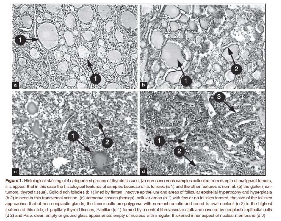
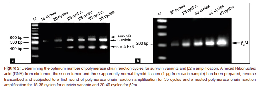
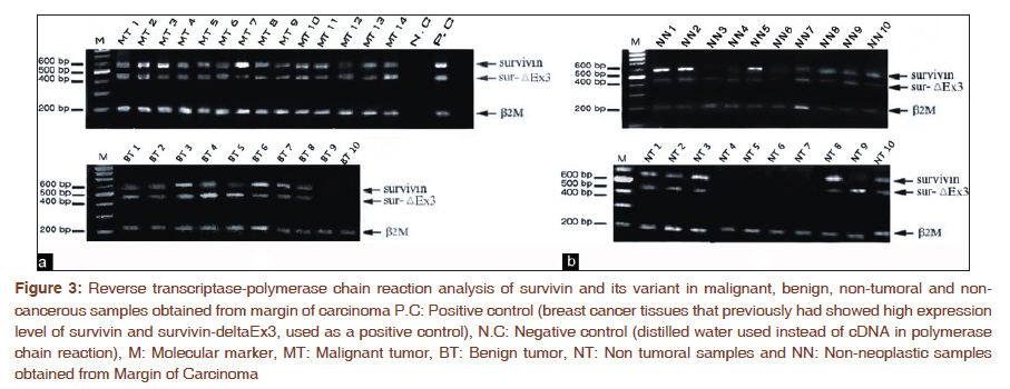
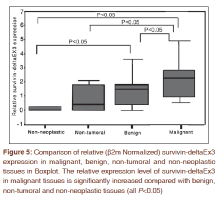
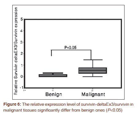
Journal of Cancer Research and Therapeutics, Vol. 7, No. 3, July-September, 2011, pp. 325-330 Original Article Survivin-deltaEx3: A novel biomarker for diagnosis of papillary thyroid carcinoma Somayeh Vandghanooni1, Morteza Eskandani2, Vahid Montazeri3, Monireh Halimi3, Esmaeil Babaei4, Mohamad Ali Hosseinpour Feizi5 1 Department of Biology-Genetics, University of Tabriz; Research Center for Pharmaceutical Nanotechnology, Student Research Committee, Tabriz University of Medical Sciences, Tabriz, Iran, PMID: 22044815 DOI: 10.4103/0973-1482.87038 Aim: To semi-quantitative detection of survivin and its splice variant, survivin-deltaEx3, in thyroid nodules. Setting and Design: We evaluated the expression level of mentioned biomarkers in thyroid nodules including carcinoma. Materials and Methods: Samples were collected from 61 thyroid nodules including malignant, adenoma, non-tumoral (goiter and thyroidities) as well as non-neoplastic normal tissues. Transcriptional levels were measured using semi-quantitative reverse transcriptase-polymerase chain reaction (RT-PCR) and the results were normalized to b2microglubin (b2m) gene. Statistical Analysis Used: Independent sample t-test was used to assess the significant variation of expression between different groups. Result: Our data for a first time revealed that survivin-deltaEx3 is significantly up-regulated from normal to malignant thyroid carcinoma tissues (approximately ten fold). Conclusion: High expression level of survivin and survivin-deltaEx3 in malignant papillary thyroid carcinoma suggested survivin gene expression and its splice variant, survivin-deltaEx3, can be potential new markers in diagnosis of human papillary thyroid carcinoma. Keywords: Malignant carcinoma, reverse transcriptase-polymerase chain reaction, semi quantitative polymerase chain reaction, splice variant, survivin Introduction Thyroid cancer, the most common type of endocrine malignancy, accounts for the majority of endocrine cancer related to death each year. [1] Well-differentiated thyroid cancers arise from follicular cells and encompasses papillary, follicular and hurthle carcinomas. Other histological types of thyroid cancer are medullary and anaplastic. [2],[3] Papillary thyroid cancer is the most common type of thyroid tumors, making up about 70-80% of all thyroid cancers, and can occur at any age. [4] Involvement of the lymph node is relatively common in papillary carcinoma and lymphatic spread is the major target of the metastasis. [5] Because of the heterogeneous nature of tumoral and non-tumoral thyroid nodules as well as the lack of suitable clinico-pathological parameters, diagnostic criteria for thyroid cancer are highly variable. [6] Therefore, finding any appropriate molecular marker involved in thyroid cancer will be useful in exact diagnosis of malignant tumors, especially managing appropriate treatments. [7],[8] As known, derangement in apoptosis would be involved in tumorigenesis. [9] Apoptosis has been identified as an intricate and critical mechanism for active cell elimination and tissue homeostasis. Defects in apoptosis pathway may lead to uncontrolled cell proliferation and tumorigenesis. Therefore, inhibitors of apoptosis can be involved in tumorigenesis and cancer progression. [10] Survivin is a member of the inhibitor of apoptosis protein family (IAPs) that has attracted attentions from several viewpoints of research. It plays a key role in the regulation of apoptosis and cell division. Five different variants are produced from survivin pre-mRNA during alternative splicing process. In addition to wild type of survivin, two survivin isoforms (i.e., survivin-2B and survivin-deltaEx3) are generated by insertion of an alternative exon 2 and removal of exon 3, respectively. [11],[12],[13] The other isoforms have been identified as survivin-3B and survivin-2a. [14] Survivin is abundantly expressed both during normal fetal development and in a broad spectrum of human cancers but is barely detectable in normal well differentiated tissues. [11] The differential expression of survivin in some types of cancers versus normal differentiated tissues makes it a useful molecular marker in cancer diagnosis and a promising therapeutic target. [15],[16],[17] In the present study, the expression level of survivin and its splice variant, survivin-deltaEx3, were evaluated in 61 specimens classified in 4 groups; papillary thyroid carcinoma, benign tumors, non-tumoral nodules including goiter and thyroidities and non-neoplastic samples obtained from margin of carcinoma specimens. However the aim of this study is investigation of the expression levels of survivin and Survivin-deltaEx3 in thyroid nodules and evaluation of their value as diagnostic markers in papillary thyroid carcinomas. Materials and Methods The research was conducted in accordance with the Helsinki Declaration and research was approved by the ethics committee of hospital and the University of Tabriz. Moreover, informed consent was obtained from all patients undergoing surgery. All the specimens were obtained in surgical operation from Imam Khomeini hospital, Tabriz, Iran. Biopsies were snap frozen in liquid nitrogen and stored at -80°C until required for Ribonucleic acid (RNA) extraction. The histopathological analyses were performed according to the World Health Organization [18] and tissue samples were classified as malignant tumors, benign tumors, non-tumoral nodules including goiter and thyroiditis and finally adjacent non-neoplastic tissues for obtaining normal tissues from each patients [Figure - 1]. Clinico-pathological diagnosis of malignant papillary carcinomas was based on the some features of the cells such as neoplasticity with glass appearance nuclear and papillae formation. For RT-PCR reaction, total RNA was extracted with the RNeasy Micro Kit (Qiagen, Hilden, Germany) exactly according to the manufacturer′s instructions. However, the integrity of the extracted RNA was evaluated by agarose gel electrophoresis and purity of RNA was examined by optical density measurement (A260/A280 ratio). The same amount of each extracted RNA sample (5 μg) was used for cDNA synthesis using Oligo-dT primers and revert Aid TM MMulv Reverse transcriptase (fermentase co. Lituania). Thereafter, two rounds of PCR reaction were performed to amplify survivincDNA (accession number: NM-001168). First round of PCR was performed in a final volume of 50μl on Techne thermal cycler using HFP 5΄-TGGCAGCCCTTTCTCAAG-3΄ (as forward primer) and HRP 5΄- GAAGAAACACTG GGCCAA G-3΄ (as reverse primer). The reaction was performed as initial denaturation at 94°C for 120 s, followed by 35 cycles denaturation at 94°C for 30 secs, annealing for at 57°C for 30secs, extension at 72°C for 60secs and a final extension at 72°C for 5 mins. Second round of PCR was performed similar to the one described for the first round except for using 28 cycles of reactions as well as internal: HFPN 5΄-ACCACCGCATCTCTACATTC-3΄ (as forward primer) and HRPN 5΄-GTTCCTCTA TGGGGTCGTC-3΄ (as reverse primer). These primers amplified 556bp and 438bp segments for survivin and surviving-deltaEx3, respectively. b2m (accession number: NM_004048) was amplified with identical conditions of first round of PCR of survivin except for using specific primers including: HBF 5΄-CTACTCTCTCTTTCTGGCCTG-3΄ (as forward primer) and HBR 5΄-GACAAGTCTGAATGCTCCAC-3΄ (as reverse primer), as well as 30 cycles of reaction. These primers amplified a 191bp segment from b2m cDNA. In this study, to quantifying PCR reaction, products were separated on a 1.5% agarose gel and visualized by ethidium bromide staining. The amount of DNA was quantized by measuring the intensity of light emitted from corresponding bands under UV light using Labimage software (version 2.6; Kapelan GmbH Co., Germany). To ensure that equal amounts of RNA were used for each reaction and those potential differences in signal intensity were not due to differences in the amounts of starting RNA, b2m was used as an internal control for each reaction, RT-PCR was performed in separate tubes under similar conditions (except for the cycle number) for survivin, survivin-deltaEx3 and b2m with results expressed as Survivin/b2m and survivin-deltaEx3/b2m expression ratios, Also, amplified products from all sample sets were loaded onto a single agarose gel and electrophoresed. Finally, pictures were then captured under identical brightness/contrast conditions, thus maximizing the accuracy of quantification. To further confirm the accuracy of the Survivin PCR products, the amplification products were purified from the gel with a kit (Qiagen, Hilden, Germany) and sequenced on both forward and reverse direction and analyzed on a DNA sequencer (Model 3100 sequencer; ABI) according to the manufacture′s specifications, with the intermediately Neday e Fan company in Iran and matched with the NCBI survivin mRNA sequence (NM-001168). Finally, all experiments were replicated three times and the semi quantitative numerical results were analyzed by performing independent sample t-test using SPSS 15, with P<0.05 considered as statistically significant. Results In order to perform semi quantitative RT-PCR and prevent PCR reaction reaches to the stationary phase and optimization of RT-PCR, the number of cycles for PCR for b2m and survivin were optimized. It was found that the appropriate cycle for amplifying b2m was 30. Also 28 cycles were used to amplify survivin and its splice variant in second round of PCR [Figure - 2]. The expression level of survivin and its splice variant, survivin-deltaEx3, in tumoral, non tumoral and non-neoplastic samples adjacent carcinoma lesion were detected by RT-PCR. [Figure - 3] showed that upper bands are survivin and Survivin-deltaEx3 respectively and the lower bands are a segment of b2m which was used as an internal control gene. Our data showed that the expression level of survivin was increased from normal tissues to malignant tumor samples. It was detected in 76% of malignant tumor, 54.5% of benign tumor, 36.6% of non tumoral and 21.4% of adjacent non-neoplastic tissues. The expression level of Survivin-deltaEx3 in malignant tumor samples was more than the other groups and detected in 84% of samples compared 36.6% of benign and non-tumoral samples. In addition its expression in adjacent non-neoplastic tissues group was apparently lower than others and was detectable in 7.14% of specimens. Also in this investigation the relative expression levels of survivin/b2m and survivin-deltaEx3/b2m for each sample were performed and compared between different groups using independent sample t- test. Results showed that the expression level of survivin in tumoral samples was significantly higher than the adjacent non-neoplastic tissues and non tumoral ones [Figure - 4] (P<0.05). In addition, there was a significant increase in transcriptional level of Survivin-deltaEx3 in tumoral tissues compared with adjacent non-neoplastic tissues [Figure - 4]. The fantastic result for this research is the higher rate of survivin-deltaEx3/survivin in malignant carcinoma compared with benign ones. Discussion In the present study, we analyzed the expression level of survivin and its splice variant, survivin-deltaEx3, in tumoral, non-tumoral and adjacent non-neoplastic tissues using by semi quantitative RT-PCR technique. In previous reports on the expression of the survivin in thyroid tumors, it was revealed that the high expression of survivin was correlated with thyroid tumors, [19],[20] but a comprehensive data about the expressional level of survivin and its splice variants in tumoral and non tumoral thyroid nodules has been very rare until now. In this study we performed semi quantitative analysis on Survivin-deltaEx3 for a first time as a possible new molecular marker in thyroid tumors. Survivin has an important role in cell cycle and support the cell dividing machinery; therefore high expression level of survivin in cancer cells may overcome cell cycle checkpoints to facilitate aberrant progression of transformed cells through mitosis. [21] Anti apoptotic properties of survivin enhance the viability of cancer cells. [13] It has been reported that the survivin and its splice variants are highly expressed in several human carcinoma including lung, breast, colon and oesophageal compared with normal tissues. [22],[23],[24],[25],[26],[27],[28] Moreover Ito et al., reported the high expression level of survivin in human thyroid tumors using immunohistochemistry technique. [19] Our results showed a significantly high expression level of survivin in malignant papillary thyroid carcinoma compared with others (all remaining three groups (P<0.05). In current study, we also found a significant increased expression level of survivin-deltaEx3 in malignant tumors compared with adjacent non-neoplastic tissues (P<0.05) and non-tumoral ones (P<0.05) [Figure - 5]. We saw a few differences in expression level of survivin-deltaEx3 in benign tumors versus non-tumoral ones, but it was not significant (P=0.526). Also, our data showed that the expression of survivin-deltaEx3 was significantly elevated in malignant tumor tissues compared with benign tumors (P<0.05) [Figure - 5]. In current study, the rate of expression level of survivin-deltaEx3 in studied groups (Malignant>benign >non-tumoral >adjacent non-neoplastic tissues) showed that this variant has conserved its anti-apoptotic properties in concordance with result of mohatka et al. [29] Furthermore, the high rate of survivin-deltaEx3/survivin in malignancy compared to benign [Figure - 6] show that in the malignant carcinoma the splicing process of survivin is highly carried out to obtain survivin-deltaEx3, so this variant can be used as a novel molecular marker to distinct between benign and malignant thyroid cancer. Additionally, the most important and annoying factor in the recognition of different types of thyroid cancer is the diagnosis of follicular carcinoma from others. The next step of this study is definitely to investigate the expression of survivin and its splice variant in follicular carcinoma and adenoma. Furthermore, very well-differentiated carcinoma that is difficult to discriminate with benign tumor should be under investigation in future studies. However, further confirmatory and quantitative examination is required to establish this statement. Conclusion In conclusion, the current study demonstrated that evaluating survivin gene expression might have a potential usefulness in diagnosis and classification of thyroid tumors from non-tumoral ones as well as introduced survivin-deltaEx3 as a new potential tumor marker in diagnosis of malignant tumors from benign ones. However, the fact that confirmatory and protein level studies of survivin and survivin-deltaEx3 were not performed in this investigation is the truth weakness of this study, so any more techniques such as immunohistochemistry or western blot techniques are offered for future works to confirm our data. References
Copyright 2011 - Journal of Cancer Research and Therapeutics The following images related to this document are available:Photo images[cr11079f3.jpg] [cr11079f4.jpg] [cr11079f5.jpg] [cr11079f2.jpg] [cr11079f6.jpg] [cr11079f1.jpg] |
| |||||||||
