 |
| About Bioline | All Journals | Testimonials | Membership | News |
|
||||||
|
||||||
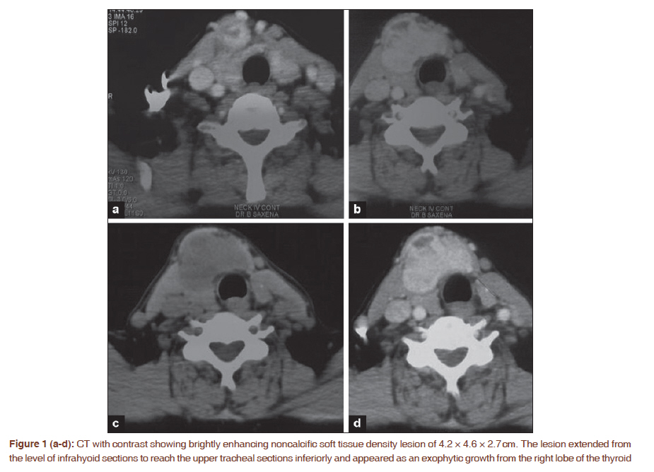
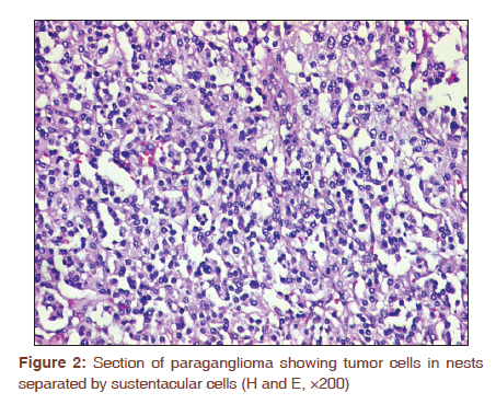
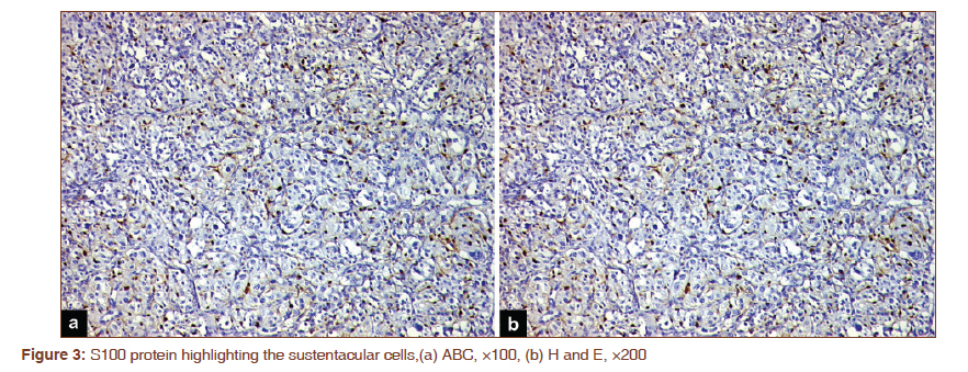
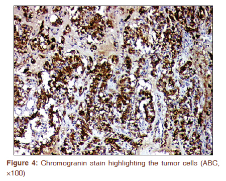
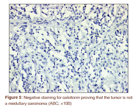
Journal of Cancer Research and Therapeutics, Vol. 7, No. 3, July-September, 2011, pp. 385-387 Letter to the Editor - Documenting a Case Primary paraganglioma of thyroid presenting as solitary thyroid mass Sandip Basu1, Seethalakshmi Viswanathan2 1 Radiation Medicine Centre, Bhabha Atomic Research Centre, Tata Memorial Centre Annexe, Parel, Mumbai, India Correspondence Address: Code Number: cr11101 PMID: 22044837 DOI: 10.4103/0973-1482.87028 Sir, We herein report a case of primary thyroid paraganglioma, an extremely rare clinical entity, with follow-up data of 1 year after surgery. The patient, a 70-year-old woman presented with a solitary thyroid nodule involving the right lobe of thyroid and isthmus of around 3.5 × 1.5 cm in dimension. On ultrasonography (USG) of the neck, the nodule appeared well-defined hyperechoic and solid and demonstrated increased vascularity on color Doppler USG. Computed tomography with contrast [Figure - 1]a-d revealed a brightly enhancing noncalcific soft tissue density lesion of 4.2 × 4.6 × 2.7 cm extending from the level of infrahyoid sections to reach the upper tracheal sections inferiorly, appearing as an exophytic growth from thyroid. She had undergone complete excision of the thyroid nodule. On cut section, the tissue was uniformly grey brown. Sections showed a well-circumscribed neoplasm with rich vascularity and focal organoid Zell-Ballen pattern [Figure - 2]. On immunohistochemistry, S100 had highlighted sustentacular cells [Figure - 3]a and b. Tumor cells were positive for chromogranin [Figure - 4], NSE, CD56, focally for synaptophysin and negative for calcitonin [Figure - 5]. Overall features were consistent with primary thyroid paraganglioma. She was not subjected to further surgery and followed-up based on age and associated cardiac morbidity. At 1-year follow-up, the patient is alive without evidence of recurrence elsewhere in the body. A whole body 131 I-MIBG scan was within normal limits 6 months after surgery and the 24 h urinary VMA level was also normal at that time. Primary thyroid paraganglioma is a rare entity with around 25 cases reported in the literature till date. [1] Overall it runs a benign course with complete removal of the disease after surgery. It is also important to know that paraganglioma is usually detected as a hypoechoic thyroid nodule on USG, although in this case the nodule appeared hyperechoic. In a systematic review published recently, there was no evidence of metastases in the reported cases and was associated with bilateral or monolateral carotid body tumors in some cases. [2],[3] Surgery in the form of hemithyroidectomy appears adequate with long-term follow-up. External radiation therapy is advocated only when there is suspicion of inadequate removal of the disease. References
Copyright 2011 - Journal of Cancer Research and Therapeutics The following images related to this document are available:Photo images[cr11101f2.jpg] [cr11101f5.jpg] [cr11101f4.jpg] [cr11101f3.jpg] [cr11101f1.jpg] |
| |||||||||
