 |
| About Bioline | All Journals | Testimonials | Membership | News |
|
||||||
|
||||||
Indian Journal of Dermatology, Venereology & Leprology, Vol. 69, No. 3, May-June, 2003, pp. 239-240 Case Report Localized cutaneous sporotrichosis lasting for 10 years Sanjay K. Rathi, M. Ramam,* C. Rajendran** *Associate Professor, Departmant of Dermatology and Venereology, All India
Institute of Medical Sciences, New Delhi, India; and
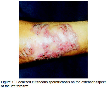
Code Number: dv03016 Abstract A case of localized cutaneous sporotrichosis lasting for 10 years is being reported. The fixed cutaneous variety creates diagnostic difficulty by mimicking other conditions, chiefly lupus vulgaris. Key Words: Sporotrichosis, Lupus vulgaris Introduction Sporotrichosis is a fungal infection caused by Sporothrix schenckii.1 Both cutaneous and systemic forms of sporotrichosis exist. Three clinical types are described: (i) fixed cutaneous, (ii) lymphocutaneous, and (iii) disseminated. Lymphocutaneous sporotrichosis is easy to recognize but fixed sporotrichosis can pose diagnostic problems.2,3 The following report illustrates the diagnostic problem caused by the fixed type of cutaneous sporotrichosis mimicking lupus vulgaris. Case Report A 60-year-old woman presented with a single, well-defined, erythematous, mildly scaly plaque with central clearing and scarring on the extensor of the left forearm (Figure 1). Thickened lymphatic cords were not palpable in the vicinity nor was there any significant lymphadenopathy. The plaque began as a small papule 10 years earlier. It gradually increased in size for the first 2-3 years and had remained static since then. There was itching and superficial ulceration of the plaque off and on. She did not recall any history of thorn prick or any other injury at the site of the lesion. There was no history of cough, hemoptysis or weight loss. A biopsy had revealed granulomatous inflammation and she was treated with three antitubercular drugs for six months. However, there was no improvement with this treatment. Her general physical and systemic examinations were normal. Her routine laboratory parameters, including the liver and renal function tests, and X-ray chest, were within normal limits. HBsAg, HCV and Mantoux test were negative. Ultrasonography of the abdomen was normal except for fatty infiltration of liver. A second skin biopsy showed extensive mixed cell granulomas in the upper and mid dermis with neutrophilic abscesses and many eosinophils. No organisms were seen. Tissue sent for fungal culture grew Sporothrix schenckii. The patient was treated with a saturated solution of potassium iodide orally, with the dose gradually increased to 45 drops 3 times a day. The lesion resolved completely with scarring after 8-9 weeks of treatment. Potassium iodide was continued for two months after clinical resolution and then stopped. The patient did not report any adverse effects. There was no evidence of disease activity when she was reviewed one and half years after completing therapy. Discussion The lymphocutaneous variety of sporotrichosis presents a distinctive clinical picture, with nodules and ulcers arranged linearly along the lymphatics with thickened lymphatic cords between the nodules, usually on exposed skin.1,3 The fixed variety, where the pathogen remains localized, is less common.4,5 It may be a nodular, acneiform, ulcerated or verrucous form of variable duration.6 The fixed cutaneous type is more difficult to diagnose and may be mistaken for other causes of a persistent, non-healing ulcer or nodule, chiefly tuberculosis.6-8 Many patients, like our case, are treated for cutaneous tuberculosis. In our patient, the diagnosis of sporotrichosis was suspected by the finding of a mixed cell granuloma on biopsy and confirmed by fungal culture. She responded well to treatment with potassium iodide. References
Copyright 2003 - Indian Journal of Dermatology, Venereology & Leprology. Free full text also available from: http://www.ijdvl.com The following images related to this document are available:Photo images[dv03016f1.jpg] |
| |||||||||
