 |
| About Bioline | All Journals | Testimonials | Membership | News |
|
||||||
|
||||||
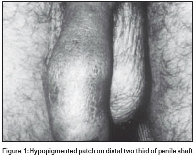
Indian Journal of Dermatology, Venereology, Leprology, Vol. 69, No. 6,Nov-Dec , 2003, pp. 411-412 Case Report Penile shaft lesion in reactional borderline tuberculoid leprosy: A case report Ghorpade A Department of Dermatology, Venereology & Leprology, J. L. N. Hospital & Research Center, Bhilai, Chhattisgarh Code Number: dv03079 ABSTRACT A twenty-year-old man with borderline tuberculoid leprosy with lesion on the penile shaft is reported. The lesion was detected when the patient developed an upgrading reaction after MDT. The diagnosis was confirmed by histopathology. Leprosy lesions are uncommonly reported on the penis and interestingly, some seem to have been detected during reactional episodes only. It appears that like facial lesions, genital patches may also be more prone to develop a type 1 reaction. INTRODUCTION Genital leprosy lesions may remain undetected in many patients, unless specifically looked for.[1],[2] They have been mostly observed in multibacillary cases having extra-genital patches too. Exclusive penile shaft involvement in tuberculoid leprosy has been reported only once.[3] CASE REPORT A 20-year-old unmarried man presented with a history of reddish skin patches on his back, left leg and over the penis since 5 months. He was taking PB MDT for BT leprosy from a private practitioner for the previous two months. He had not shown the penile lesion to the doctor, who did not also specifically look for genital lesions. There was a sudden swelling in the existing skin patches and inability to retract the foreskin for the past three weeks. He did not develop any fresh lesions. There was no history of burning micturition, sexual contact, fever or joint pains. His family members did not have any skin problem. Cutaneous examination revealed four circular, well defined, erythematous, edematous, dry, anesthetic plaques on his back and one on the lower part of his left leg on its lateral aspect. The lesions varied from 5 cm to 17 cm in diameter. A similar shiny, erythematous, edematous, hypoesthetic, warm plaque of about 4 cm x 3.5 cm was seen on the dorsal and lateral aspects of the distal two-thirds of the penile shaft [Figure - 1], extending up to the tip of the prepuce. There was edema of the distal half of the penile shaft and prepuce with partial phimosis. The glans penis and scrotum were free from lesions. The patient was afebrile and did not have edema of the hands and feet. The left lateral popliteal nerve was thickened and tender. Slit skin smear examination from the routine sites and the penile plaque for M. leprae was negative. His systemic examination and routine hematology were normal. The blood VDRL was negative. Histopathology from the penile lesion revealed multiple compact upper dermal granulomas of epithelioid cells, lymphocytes and Langhans′ giant cells. There was edema in the upper epidermis. Ziehl-Neelsen stains of the section did not show any AFB. He was diagnosed as a case of borderline tuberculoid leprosy with upgrading reaction. Prednisolone 30 mg/day was added to the PB MDT. It was tapered over a 4 month period, during which the edema regressed, while the PB MDT was continued for 11 months, after which the skin lesions almost cleared. DISCUSSION Genital lesions in leprosy have not been frequently reported and may be missed. Arora et al[1] and Kumar et al[2] reported genital lesions in 2.9% and 6.6% of leprosy patients respectively. Most of their cases belonged to the borderline and borderline lepromatous group respectively. Genital lesions have been rarely reported in histoid leprosy.[2],[4],[5] Scrotal involvement has been commonly observed along with penile lesions.[1],[2],[6] Pandya and Antia found leprous granulomas and AFB in biopsies from apparently normal scrotal skin.[7] Upgrading reactional episodes of leprosy, occurring in penile leprosy lesions, as in our patient, have been observed earlier. Of the 13 cases of external genital involvement reported by Arora et al, most were in type 1 reaction.[1] Parikh et al found a type 1 reaction in two out of six patients with genital lesions.[6] In another case, the leprosy lesion on the prepuce of a borderline leprosy patient was noticed after he developed a type 1 reaction while on MDT.[8] Why the genital leprosy lesions undergo reversal reactions frequently is difficult to explain. It is also interesting to note that the penile lesions were found on the distal two-thirds of the penile shaft, as in earlier patients.[3],[8] Is it that the proximal one-third of the penile shaft is comparatively immune, probably due to a slightly higher temperature? The predilection of facial lesions to undergo type 1 reaction after MDT is well known. The present case and the earlier reports suggest that, like facial lesions, the penile lesions too may be more prone to develop a leprosy reaction. A detailed study of genital leprosy lesions might uncover the probable reasons. REFERENCES
Copyright 2003 - Indian Journal of Dermatology, Venereology, Leprology The following images related to this document are available:Photo images[dv03079f1.jpg] |
| |||||||||
