 |
| About Bioline | All Journals | Testimonials | Membership | News |
|
||||||
|
||||||
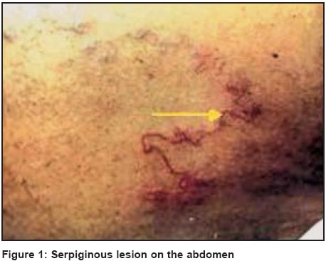
Indian Journal of Dermatology, Venereology, Leprology, Vol. 70, No. 1, January-February, 2004, pp. 59-60 Quiz Serpiginous lesion on the abdomen Padmavathy L, Rao LLakshmana Urban Health Center, Department of Community Medicine, Rajah Muthiah Medical College, Annamalai University, Annamalai Nagar - 608 002 Code Number: dv04020 A 60-year-old retired teacher presented with an intensely pruritic, progressive, serpiginous lesion of six weeks′ duration on the anterior abdominal wall. Gardening was his hobby. He gave a history of contact with the soil while changing the mud in pots and while manuring plants. He had a dog as a pet. Clinical examination revealed a curvilinear, erythematous lesion about 20 cm long on the anterior abdominal wall, healing at one end and progressing at the other [Figure - 1]. Baseline hematological and biochemical investigations were within normal limits. What is your diagnosis? diagnosis: creeping eruptionTreatment with oral albendazole and topical cryotherapy resulted in clearance of the lesion in 4 weeks. The pet dog was also de-wormed. Creeping eruption is a distinctive condition due to numerous etiological agents, like Ankylostoma brazilienses, A. caninum, Uncinaria stenocephala, Gnathostoma sp., Dirofilaria conjunctivae, Capillaria species, etc.[1] Other synonyms of this condition are "cutaneous larva migrans", "sand worms", creeping verminous dermatitis, plumber′s itch and duck hunter′s itch. Creeping eruption commonly occurs when the larvae of dog or cat hookworms penetrate intact exposed skin and migrate though the epidermis. Lacking the enzymes necessary to penetrate and survive in the deeper dermis, the larvae wander in a serpiginous route at a speed of 3 cm/day and produce the typical lesion.[2] The incubation period ranges from 1 to 6 days. The most common location for the eruption is the feet, but other sites like buttocks, back, and thighs may also be affected. Larva migrans over the genitalia,[3] oral mucosa,[4] and in a 6-month-old infant[5] have been reported. Though the condition is self limited, secondary bacterial infection and eczematization can occur. Extensive lesions may be associated with wheezing, dry cough and urticaria. A. caninum larvae can migrate to the small intestine and cause eosinophilc enteritis. Transient eosinophilia has been also reported.[5] The diagnosis is established by the classic clinical picture. Biopsy is of no utility as the larvae advance ahead of the clinical tract. Epiluminiscence microscopy is an effective non-invasive method to detect the larva and confirm the diagnosis.[6] The lesions disappear in 2-8 weeks but rarely may persist for 2 years. Useful drugs include topical and oral thiabendazole, mebendazole and ivermectin (150 to 200 mcg/kg as a single dose).[7] REFERENCES
Copyright 2004 - Indian Journal of Dermatology, Venereology, Leprology The following images related to this document are available:Photo images[dv04020f1.jpg] |
| |||||||||
