 |
| About Bioline | All Journals | Testimonials | Membership | News |
|
||||||
|
||||||
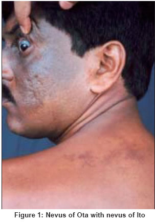
Indian Journal of Dermatology, Venereology, Leprology, Vol. 70, No. 2, March-April, 2004, pp. 112-113 Case Report Nevus of ota associated with nevus of Ito Mukhopadhyay Amiya Kumar Consultant Dermatologist, Asansol Code Number: dv04034 ABSTRACT Nevus of Ota is a dermal melanocytic nevus seen predominantly in females. It is uncommon in India. Its association with nevus of Ito, another dermal melanocytic nevus, is extremely rare. We report this rare association in a male patient, which is another interesting feature of the present case.INTRODUCTION First described by Ota in 1939, nevus of Ota is an extensive, blue patch of dermal pigmentation involving the eyelids, the bulbar and palpebral conjunctiva and sclera, the cheeks, forehead, scalp, alae nasi and ears. The mucosa of the cheeks and palate may also be affected.[1] The condition is uncommon in India. It is very rare in male patients.[2] Nevus of Ito was first described in 1954 by Ito. It has the same features of Ota′s nevus except that it occurs in the distribution of the posterior supraclavicular and lateral cutaneous branchial nerves, to involve the shoulder, side of the neck and supraclavicular areas.[3] We report the simultaneous occurrence of nevus of Ota and nevus of Ito in a male patient. CASE REPORT A 38-year-old man presented with an asymptomatic hyperpigmented lesion on the left side of the face, left eyelid and sclera of the left eye. He also had a similar lesion on the left shoulder. According to the patient, the facial and the shoulder lesions were present since childhood, but the former had appeared earlier. After gradual progression to the age of 25 years the lesions had reached the present size and then remained static. There was no significant family history On examination, bluish grey pigmentation of the left side of the face, left eye and left sclera was noted. The lesions on the shoulder were of a similar color, but diffuse in nature, with some darker macules at places [Figure - 1]. Ophthalmological examination revealed no abnormality. General examination showed no abnormality. Histopathological examination of the lesions showed dermal dendritic melanocytes scattered in the upper portion of the dermis. DISCUSSION Both nevus of Ota and nevus of Ito are dermal melanocytic nevi. They are particularly common in the Japanese.[1] Nevus of Ota affects about 0.8% of the Japanese,[4] but is uncommon in India.[2] Its distribution is usually restricted to the first and second divisions of the trigeminal nerve, but rarely lesions may occur on the trunk.[3] The present case is interesting because of the very rare association of nevus of Ota and nevus of Ito in the same patient and also because the patient is a male, whereas 80% of the cases of nevus of Ota occur in females.[2] REFERENCES
Copyright 2004 - Indian Journal of Dermatology, Venereology, Leprology The following images related to this document are available:Photo images[dv04034f1.jpg] |
| |||||||||
