 |
| About Bioline | All Journals | Testimonials | Membership | News |
|
||||||
|
||||||
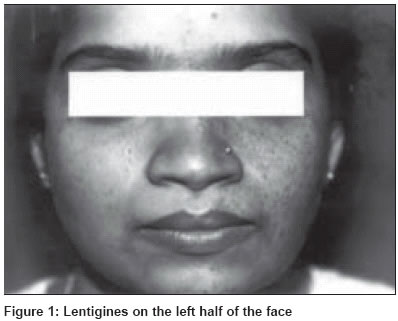
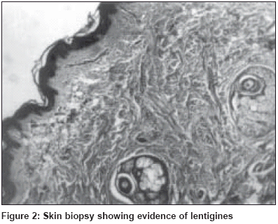
Indian Journal of Dermatology, Venereology, Leprology, Vol. 70, No. 2, March-April, 2004, pp. 114-115 Letter To Editor Partial unilateral lentiginosis with lisch nodules: A forme fruste of segmental neurofibromatosis? Rao Gatha S Department of Skin and STD, K. M. C Hospital, Attavar, Mangalore Code Number: dv04035 Sir, Partial unilateral lentiginosis is an unusual pigmentary disorder characterized by multiple lentigines in a unilateral distribution. The lesions often have a segmental pattern with a sharp demarcation at the midline. Histologically they are characterized by prominent rete ridges of the epidermis and an increase in the melanin content of the basal layer.[1] A 21-year-old female presented with multiple hyperpigmented macules on the left side of the face of 17 years′ duration. Hyperpigmented macules were noticed on the left side of the cheek at the age of 4 years, which gradually progressed to involve the left half of the face, neck, chest and upper back. No history of photosensitivity was present. There was no history of epilepsy, mental retardation or a family history of similar illness. The patient gave a history of easy fatiguability since childhood. On examination, the patient was pale. Systemic examination revealed no abnormality. Cutaneous examination showed multiple, discrete, hyperpigmented macules of sizes varying from 2-5 mm on normal skin on the left half of the face, neck, On investigation, the patient had hypochromic microcytic anemia with hemoglobin of 8.5 g%. An electrocardiogram, echocardiography, and ultrasonography of the abdomen were within normal limits. A skin biopsy showed aggregates of melanocytes with increase in melanin in the basal layer and elongation of rete ridges suggestive of lentigines [Figure - 2]. Melanin bleach was positive. The lesions of partial unilateral lentiginosis first appear during childhood. They can appear anywhere on the body but the upper extremities are more affected than the lower ones.[1] The condition results from mutation during embryonic development probably confined to neural crest melanoblasts.[2] It can be associated with various central nervous system abnormalities including mental retardation,[3] neurofibromatosis,[4] cerebrovascular abnormalities with focal epilepsy,[2] and probably with iron deficiency anemia and euthyroid goitre.[1] The coexistence of neurofibromatosis with partial unilateral lentigines raises the possibility that partial unilateral lentigines could be a variant or forme fruste of segmental neurofibromatosis.[5] Our patient had no cutaneous lesions of neurofibromatosis but had bilateral Lisch nodules in the eyes. Since Lisch nodules are characteristic of neurofibromatosis, we conclude that the partial unilateral lentiginosis seen in our patient is a forme fruste of neurofibromatosis. REFERENCES
Copyright 2004 - Indian Journal of Dermatology, Venereology, Leprology The following images related to this document are available:Photo images[dv04035f2.gif] [dv04035f1.jpg] |
| |||||||||
