 |
| About Bioline | All Journals | Testimonials | Membership | News |
|
||||||
|
||||||
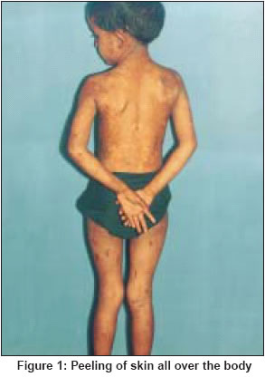
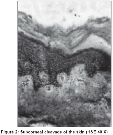
Indian Journal of Dermatology, Venereology, Leprology, Vol. 70, No. 2, March-April, 2004, pp. 115-116 Letter To Editor Peeling skin syndrome Mukhopadhyay Amiya Kumar "Pranab", Ismile (Near Dharmaraj Mandir), P.O. Asansol - 713301 Code Number: dv04036 Sir, Peeling skin syndrome (PSS) is a rare disorder characterized by continual shedding of the entire stratum corneum. It starts either at birth or later in childhood.[1] It is inherited in an autosomal recessive pattern.[2] The skin involvement is usually generalized, but in some patients sparing of the face, palms and soles has been reported.[3] Here we report a case of PSS in a girl who had sparing of palms and soles and whose disease showed summer exacerbation. A 5-year-old girl presented with a history of peeling of skin from all over the body in patches since 1 year of age. She was born of a non-consanguineous marriage and there was no history of a similar disorder in any of her family members. Her problem of peeling of the skin remained all around the year but worsened in the summer. The lesions were mildly itchy. On examination she had patches of peeling skin all over the body along with a few lesions on her face [Figure - 1]. Rubbing the normal skin revealed the peeling but there was no oozing from the rubbed area. Some hyperpigmented patches remained at the sites of old lesions. The palms, soles and mucous membranes were uninvolved. General and systemic examination revealed no abnormality. Routine hemogram and urinalysis were normal. Histopathological examination of the skin showed separation of the stratum corneum away from the stratum granulosum, mild acanthosis and a normal dermis [Figure - 2]. The patient was treated with emollients which gave her symptomatic relief to some extent, but on follow up she had recurrence of the lesions. PSS is variously known as keratolysis exfoliativa, congenital deciduous skin, and familial continual skin peeling.[4] Troupe has pointed out that there are two types of peeling skin syndrome. The first was first described by Fox in 1921, and is characterized by shedding of the entire skin including the face, comparable to the shedding of skin by reptiles. The peeling occurs in the lower part of the stratum corneum. The second type, first described by Wile in 1924, presents with erythroderma at birth, and is associated with features such as growth retardation, aminoaciduria, etc. The epidermis is psoriasiform with an absent or reduced granular layer and with marked parakeratosis. The split occurs at the level of the granular layer.[2] It has been suggested that the basic lesion is located at the stratum corneum-granular layer junction; ultrastructually, intercellular disruption occurs above the lower two layers of the stratum corneum within the stratum lucidum.[4] The cause of this disorder is unknown.[5] It has been postulated that the defect is reduced adherence of abnormally thick stratum corneum to the stratum granulosum.[6] In some patients, association of easy pluckability of hair, shedding of nails, hypogonadism and anosmia has been described.[4] Usually there are no seasonal exacerbations, but some patients appear to worsen in summer.[5] As far as treatment is concerned, emollients such as petrolatum jelly and salicylic acid gel may be helpful in improving the appearance.[7] Drugs derived from vitamin A, such as tretinoin and etretinate, could be effective.[8] In the present case, the history of exacerbation during the summer and sparing of the palms and soles are uncommon and interesting features. REFERENCES
Copyright 2004 - Indian Journal of Dermatology, Venereology, Leprology The following images related to this document are available:Photo images[dv04036f1.jpg] [dv04036f2.jpg] |
| |||||||||
