 |
| About Bioline | All Journals | Testimonials | Membership | News |
|
||||||
|
||||||
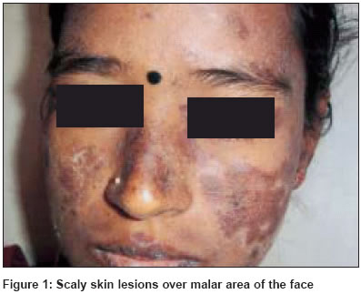
Indian Journal of Dermatology, Venereology, Leprology, Vol. 70, No. 4, July-August, 2004, pp. 243-244 Letter To Editor ANA-negative systemic lupus erythematosus Pratap DVS, Reddy SivaramiK, Rani SudhaC, Krishna VenkataA, Indira D Department of Dermatology, Gandhi Medical College, Hyderabad Code Number: dv04081 Sir, We report a case of ANA-negative systemic lupus erythematosus diagnosed on the basis of ARA criteria. A 27-year-old female presented with fever and bilaterally symmetrical dusky erythematous, scaly papules and patches with pigmentation over the scalp, malar area of the face [Figure - 1], back, neck and extremities of 2 months′ duration. Her oral cavity showed non-indurated ulcers. Diffuse and patchy alopecia was observed over the parietal and occipital areas of the scalp. Investigations showed normocytic, hypochromic anemia, leukocytosis, raised ESR, hematuria, pyuria, and raised levels of total serum bilirubin, serum glutamate pyruvate transaminase and serum alkaline phosphatase. An X-ray chest showed right mid-zone consolidation. Ultrasonography of the abdomen revealed hepatomegaly suggestive of a diffuse parenchymal affection, upper abdominal lymphadenopathy with minimal ascites, and changes suggestive of acute renal parenchymatous disease. Serum HBsAg, Elisa for HIV-1 and HIV -2, serum antinuclear antibody (ANA), anti ds-DNA and LE cell tests were negative. Serum anti-Ro test was positive. The serum C3 was 94.3 mg/dl. A skin biopsy showed focal atrophy with keratotic plugging in the epidermis, basal cell liquefaction degeneration with colloid bodies, patchy mononuclear infiltrates in the dermis, and melanin incontinence. The presence of ANA is one of the criteria for the diagnosis of SLE. In 5-10% of cases of SLE, ANA cannot be demonstrated although the other ARA criteria are fulfilled.[1] About 10% of these cases may eventually become ANA positive.[1] Immunofluorescence (IF) assay or enzyme immunoassay (EIA) can detect ANA. In the IF assays substrates such as Hep2 cells, a human laryngeal cell line, are used. They give a higher incidence of positive results.[1] However, in view of the drawbacks like difficulty of reproducibility and requirement for higher quality control, EIA is a better method for routine use. In our patient, we could eventually detect ANA by using EIA designed by Bio-Rad Laboratories. This is a qualitative immunoassay using tetra methyl benzidine in dilute hydrogen peroxide buffer as substrate. The test was repeated in a couple of other laboratories using the same kit. Circulating antibodies to DNA are almost always present in active disease, and may occur in the absence of antinuclear factor.[1] Antibodies to soluble cellular antigens include anti-Sm antibody, found in 15-25% of patients with SLE, particularly in patients with renal involvement, CNS disease and vasculitis. Anti-RNP antibody occurs in 25% of patients with characteristics of mixed connective tissue disease. Anti-Ro antibody occurs in 30% of patients who will have increased tendency to photosensitivity, renal disease or Sjögren′s syndrome.[1] Anti-Ro antibody is also found in patients with subacute cutaneous lupus erythematosus (SCLE). In one Indian study, all 7 patients of SCLE were ANA-negative.[2] The presence of 6 ARA criteria in our patient, along with classical histopathological findings in the skin biopsy, strongly suggest the diagnosis of SLE in spite of the absence of ANA. Hence this case can be labeled as ANA-negative SLE. REFERENCES
Copyright 2004 - Indian Journal of Dermatology, Venereology, Leprology The following images related to this document are available:Photo images[dv04081f1.jpg] |
| |||||||||
