 |
| About Bioline | All Journals | Testimonials | Membership | News |
|
||||||
|
||||||
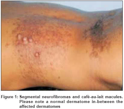
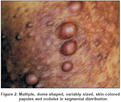
Indian Journal of Dermatology, Venereology, Leprology, Vol. 70, No. 6, November-December, 2004, pp. 361-363 Case Report Multi-segmental neurofibromatosis Kumar Sudhir, Kumar RaviP Department of Neurological Sciences, Christian Medical College Hospital, Vellore Code Number: dv04124 ABSTRACT Neurofibromatosis (NF), one of the commonest phakomatoses, is characterized by varied clinical manifestations. Segmental NF is one of the uncommon subtypes of NF. We report a young adult presenting with asymptomatic skin lesions- neurofibromas and café-au-lait macules- over localized areas of the lower back, affecting more than one segment. None of the family members were found to have features of segmental NF. Segmental NF may be misdiagnosed as a birthmark or remain undiagnosed for long periods of time, as the patients are often asymptomatic. Moreover, the clinical features are highly variable and range from a small area of skin involvement to involvement over the entire half of the body. This variation is explained by the fact that segmental NF is thought to arise from a postzygotic NF1 gene mutation, leading to somatic mosaicism. We have also reviewed the relevant literature on this subject.KEY WORDS: Segmental neurofibromatosis, NF Type 5, Genetic mosaicism INTRODUCTION Neurofibromatosis (NF) is one of the commonest genetic neurocutaneous syndromes. NF is highly heterogeneous and at least seven types have been described, with NF Type 1 being the commonest.[1] We report a young man with features of segmental NF (NF Type 5) affecting more than one segment, which is among the uncommon subtypes of NF.[2] CASE REPORT A 36-year-old man presented with low back pain of three months duration. He had no motor, sensory or autonomic symptoms. A detailed neurological examination was normal. Examination of the skin showed multiple soft, dome-shaped, flesh-colored papules and nodules (neurofibromas) on the lower back in T8-10 dermatomes, bilaterally (predominantly on the left side) with asymmetric distribution [Figure - 1]. Café-au-lait macules (CALM) with irregular margins were seen on the same side in T12 dermatome [Figure - 2]. Examination of the rest of the skin was normal. He had no axillary freckling or Lisch nodules on the iris. Magnetic resonance imaging of the spine was normal. The patient had noted these skin lesions since the age of 22 years. He had no systemic complaints. There was no history of similar illness in any other family member. Screening of his parents and three children did not reveal any features of NF. Based on the presence of neurofibromas and CALM in a circumscribed body part, lack of systemic involvement and negative family history, a diagnosis of segmental NF was made. Back pain was attributed to mechanical causes and improved with physiotherapy. Patient was counseled and advised regular follow-up. DISCUSSION Segmental neurofibromatosis (NF) is a rare variant of NF in which skin lesions are confined to a circumscribed body segment. The affected area, however, can vary in size from a narrow strip to an area encompassing half of the body. Segmental NF is under-diagnosed, as it occurs with a clinical picture that, due to absence of symptoms and complications in most affected patients, can be ignored by the patient (who assumes it to be an odd birthmark or a harmless nodule) and passes unnoticed by the physician.[2] This is supported by the fact that though the first case of segmental NF was reported in 1937,[3] less than 150 cases have been reported till date, notwithstanding an estimated prevalence of 1 in 40,000 in the general population.[2] There is a paucity of data on this disease from India too.[4],[5],[6],[7] It occurs in all age groups, has a mean age of onset of 28 years,[8] with a slight male preponderance (male: female ratio = 1.14:1).[2] The majority of the patients are asymptomatic and seek medical attention for disfigured appearance of skin. In others, the condition is incidentally diagnosed while being examined for another problem, as happened in our patient. Exceptions include those with spinal neurofibromas[4] or lesions on major peripheral nerves, who present with pain or neurological deficits.[2] Cervical and thoracic regions are commonly involved and the disease is unilateral in the majority. Bilateral involvement may be either symmetric or asymmetric.[8] Our patient had asymmetric involvement of the lower thoracic region. Various features of NF such as CALM, axillary freckling and neurofibromas occur at a varying frequency. However, variations seem to depend upon the age of onset of symptoms. In children, pigmentary findings are commoner; CALM is seen in 75% of children[9] as compared to 25% of adults.[8] Similarly, axillary freckling is seen in 56% of children[9] as compared to 11% of adults.[8] Lisch nodules are rarely seen, even after thorough ophthalmologic examination. Segmental NF does not evolve to the generalized form. Moreover, severe disease is less likely to occur in the segmental form as compared to NF1. Systemic complications typically associated with NF1 are rare and occur in less than 10% of patients. These include learning difficulties, plexiform neurofibromas, optic pathway gliomas and pseudarthrosis.[2] Malignancies rarely occur in association with segmental NF; reported cases include peripheral nerve sheath tumours[10] and adrenal ganglioneuroma.[11] Segmental NF has been reported to occur in association with a variety of conditions, which include adenocarcinoma of colon,[12] agenesis of corpus callosum,[5] nevus sebaceus of Jadassohn,[13] partial unilateral lentiginosis,[14] and renal agenesis.[15] Segmental NF needs to be differentiated from certain similar-appearing lesions, such as epidermal nevus,[16] nevus lipomatosus cutaneous superficialis,[17] and agminated lentiginosis (a condition characterized by numerous lentigines confined to a body segment, with a sharp demarcation at the midline).[18] Segmental NF is thought to arise from a postzygotic NF1 gene mutation, leading to somatic mosaicism.[19] By FISH analysis, a whole gene deletion was shown in a percentage of fibroblasts from a café-au-lait spot in the affected skin but not from unaffected skin or blood lymphocytes. NF1 mutations in Schwann cells, but not in fibroblasts, correlate with neurofibroma formation.[20] It is thought that, if a somatic mutation occurs early enough, it will result in generalized disease, which is clinically indistinguishable from the non-mosaic form of the disease.[2] Individuals with segmental NF have a low risk of transmitting the disease to their offspring. Moreover, 93% of patients do not have a positive family history.[8] The risk of transmission, however, correlates with the extent of clinical involvement. There is no specific management strategy. However, genetic counseling should be done and the small risk of transmission to the offspring communicated. Careful follow-up is required to monitor disease progression or to detect any systemic complications that may occur in a minority. REFERENCES
Copyright 2004 - Indian Journal of Dermatology, Venereology, Leprology The following images related to this document are available:Photo images[dv04124f2.jpg] [dv04124f1.jpg] |
| |||||||||
