 |
| About Bioline | All Journals | Testimonials | Membership | News |
|
||||||
|
||||||
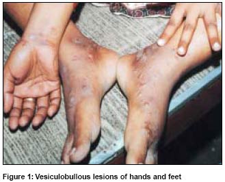
Indian Journal of Dermatology, Venereology, Leprology, Vol. 71, No. 1, January-February, 2005, pp. 53-54 Letter To Editor Bullous scabies in a patient on anticancer therapy Jena DK, Dash ML, Chhetia R Departments of Skin and VD, S.C.B. Medical College and Hospital, Cuttack Code Number: dv05018 Sir, Scabies is highly contagious and intensely pruritic skin infestation caused by mite Sarcoptes scabiei var hominis. Bullous scabies is a rare manifestation of scabies which is occasionally seen in the immunosuppressed.[1] We report a case of bullous scabies in an immunosuppressed 30 year old lady on anticancer therapy. CASE REPORT A 30 year old female patient, who was taking methotrexate, cyclophosphamide, and actinomycin D for gestational trophoblastic disease (choriocarcinoma) since 3 months, was referred for highly pruritic, vesiculobullous lesions which were present since 8 days. Itching was more severe during nights. On examination, excoriated papules and vesicles with clear fluid filled tense bullae (1-1.5 cm) were present over dorsa of feet, ventral surface of right forearm, finger webs, chest, abdomen and thighs [Figure - 1]. Closer examination revealed burrows over finger webs. Direct microscopy of scraping from the burrow revealed eggs of S. scabiei mite. Hemogram was normal. Bulla fluid on culture was sterile. Histopathology showed subepidermal bullae with cavity containing predominantly neutrophils and some eosinophils. The patient was treated with topical 5% permethrin. Resolution of the lesions was noted after 4 weeks. DISCUSSION Though the vesicular lesions are common in infants and children, bullous lesions are uncommon in scabies. Bullous scabies is seen in immunocompetent infants, young children and elderly although some reports have shown its occurrence in immunosuppressed adults.[3] In communities where scabies is not endemic, the index of suspicion is low and bullous scabies can be confused with bullous pemphigoid, insect bites, linear IgA dermatosis, epidermolysis bullosa or chronic bullous disease of childhood (CBDC). Bullous lesions over scabies prone sites, nocturnal itching and detection of mite eggs in burrow scrapping finally confirmed scabies in our case. Bullous scabies has on occasions resembled bullous pemphigoid clinically, light microscopically and immunopathologically.[1],[4] Previous case reports suggest that several cases of bullous scabies were misdiagnosed and treated as bullous pemphigoid, due to false positive DIF and IIF in bullous scabies.[4] Several theories have been proposed to explain pathogenesis of bullae formation in bullous scabies. Veraldi et al[5] has given the theory of alteration of bullous pemphigoid antigen with mite secretions and the resultant production of antibasement membrane zone antibody formation. Other theories proposed to explain bullae in scabies are superinfection with Staphylococcus aureus,[6] toxins producing bullae and id reaction to scabies mite.[7] Konishi et al[8] have demonstrated induction of bullous pemphigoid by Western Blot analysis of circulating antibodies against BP 180 and BP 230 antigen which lead them to propose that some of the lesions occurring in bullous scabies are true bullous pemphigoid. Bornhovd et al [9] postulated persistence of parasites as cause of Th2 cells activation with increased IL5 and eosinophils secretion of proteolytic enzymes near BMZ (basement membrane zone) producing bullae formation. The exact mechanism for bullae formation is not known and further studies need to be done to explain the pathogenesis of bullous scabies. In a patient with itchy, vesiculobullous lesions, burrows should be looked for, as they are seen in majority of cases of bullous scabies. REFERENCES
Copyright 2005 - Indian Journal of Dermatology, Venereology, Leprology The following images related to this document are available:Photo images[dv05018f1.jpg] |
| |||||||||
