 |
| About Bioline | All Journals | Testimonials | Membership | News |
|
||||||
|
||||||
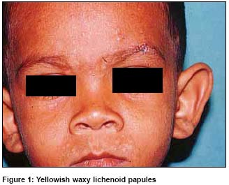
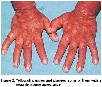
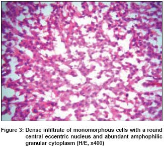
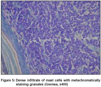
Indian Journal of Dermatology, Venereology, Leprology, Vol. 71, No. 1, January-February, 2005, pp. 63-65 Quiz Generalised yellowish papular eruption in a child Pande Sushil, Patil Rahul, Mahajan Sunanda, Kharkar Vidya, Khopkar Uday Department of Dermatology, Seth G. S. Medical College and KEM Hospital, Mumbai Code Number: dv05022 A 3 year old male child presented with multiple asymptomatic flat yellowish waxy papules and plaques over face [Figure - 1], trunk, dorsa of hands [Figure - 2] since twentieth day of life. At the age of two weeks, the child had developed generalised erythematous rash that had persisted for a few months before subsiding on its own and leaving behind the present lesions. Complete hemogram was normal. USG abdomen showed mild hepatomegaly. Skeletal survey showed no evidence of bony changes. Biopsy from the plaque revealed dense lichenoid infiltrate of monomorphous cells [Figure - 3] WHAT IS YOUR DIAGNOSIS? Diagnosis Biopsy showed a dense infiltrate of oval cells with amphophilic granular cytoplasm that stained metachromatically with Giemsa [Figure - 4] thus confirming the diagnosis of mastocytosis. In view of clinical features this case represented the rare non-pigmented, pseudoxanthomatous papulonodular form of mastocytosis which is a variant of diffuse cutaneous mastocytosis. This variety typically shows lesional erythema without urtication on stroking. DISCUSSION Other skin disorders that can present with similar pseudoxanthomatous papular lesions in a child include juvenile xanthogranuloma, xanthoma disseminatum, benign cephalic histiocytosis, generalized eruptive histiocytoma, verruciform xanthoma, eruptive syringoma, trichodiscomas, and Birt Hogg Dube syndrome (multiple fibrofolliculomas and trichodiscomas). However these can be easily differentiated from pseudoxanthomatous mastocytosis on the basis of histopathology. Cutaneous mastocytosis (CM) is the most common form of mastocytosis that predominantly affects children,[1] In a study of 71 cases of cutaneous mastocytosis in children, the most frequent clinical form of mastocytosis was urticaria pigmentosa (75%) followed by mastocytoma (17%) and diffuse cutaneous mastocytosis (8%). Darier′s sign was present in 94% of cases.[2] Diffuse cutaneous mastocytosis presents as diffuse infiltration of the skin without any discrete lesions. Entire skin is erythematous and edematous with a yellowish tinge and a peau de orange look. It later turns yellow brown to dark brown in color and is diffusely thickened and leathery due to accentuation of skin markings and flexion creases. Large blisters may develop on apparently normal skin spontaneously or following pressure or minor trauma.[3] Darier′s sign is easy to demonstrate at times with vesiculation. Pseudoxanthomatous mastocytosis is a variant of diffuse cutaneous mastocytosis described in 1875 by Tilbury. It is characterized by multiple, pale, flat yellowish papules and nodules, varying in size from 1 mm - 2 cm, present at birth and resembling xanthomatosis.[4] This multinodular nonpigmented variety of mastocytosis is extremely rare.[5] As against a classic Darier′s sign, only erythema without urtication is elicited by rubbing.[3] Skin biopsy reveals dense dermal infiltrate of mast cells which stain metachromatically with Giemsa stain. Diagnosis is confirmed on the basis of clinical features and characteristic histopathological findings. It may be apt here to remember that juvenile xanthogranuloma and mastocytosis may occur together. They are thought to share histogenesis both being reactional processes with an unspecified stimulus. [6] Treatment of pseudoxanthomatous mastocytosis is not different from other forms of cutaneous mastocytosis. Treatment options include sedative antihistamines, avoidance of trigger factors, topical or systemic steroids, mast cell stabilizers like ketotifen or disodium chromoglycate, PUVA therapy and interferons. In the absence of systemic disease, prognosis of cutaneous mastocytosis in children is generally good. In children with limited disease, most cases clear completely. Others improve or the condition persists indefinitely. Diffuse cutaneous mastocytosis in children younger than 5 years usually resolves spontaneously. REFERENCES
Copyright 2005 - Indian Journal of Dermatology, Venereology, Leprology The following images related to this document are available:Photo images[dv05022f5.jpg] [dv05022f4.jpg] [dv05022f3.jpg] [dv05022f1.jpg] [dv05022f2.jpg] |
| |||||||||
