 |
| About Bioline | All Journals | Testimonials | Membership | News |
|
||||||
|
||||||
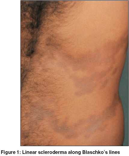
Indian Journal of Dermatology, Venereology and Leprology, Vol. 71, No. 6, November-December, 2005, pp. 421-422 Case Reports Linear scleroderma following Blaschko's lines Mukhopadhyay AmiyaKumar Consultant dermatologist, Asansol, West Bengal Code Number: dv05138 Abstract Blaschko's lines form a pattern, which many diseases are found to follow, but linear scleroderma following Blaschko's lines is a controversial entity rarely reported in the literature. A 24-year-old man presented with multiple linear, atrophic, hyperpigmented lesions punctuated by areas of depigmentations on the left half of the trunk distributed on the anterior, lateral and posterior aspects. The lesions were distributed in a typical S-shaped line. Antinuclear antibody and antihistone antibody tests were negative. Histopathological examination of the skin from the affected area showed features suggestive of scleroderma. Here, we present a case of linear scleroderma following Blaschko's lines in a male patient - an entity reported only three times so far.Keywords: Scleroderma, Linear, Lines of Blaschko Introduction Blaschko′s lines (BL) are the pattern assumed by many congenital as well as acquired disorders. They do not follow any known nervous, vascular or lymphatic distribution.[1] Localized scleroderma (LS) is divided clinically into three subclasses: (1) circumscribed plaques or band; (2) linear morphea; and (3) fronto-parietal lesions, with or without hemiatrophy of the face.[2] LS following BL is a controversial entity.[3] Of the five patients reported, only one was male. Here, we describe a male patient whose lesions were distributed along the lines of Blaschko. Case Report A 24-year-old man presented with multiple hyperpigmented, atrophic lesions on the left side of his body for the last six years. He gave a history of erythematous, indurated lesions that started spontaneously and gradually transformed into the present picture. He denied any history of trauma, disease or drug administration preceding his present problem. Other members of his family were normal. On examination, there were atrophied, hyperpigmented lesions punctuated by areas of depigmentation present on the left half of the trunk distributed on the anterior, lateral and posterior aspects [Figure - 1]. They followed a typical S-shaped line at places. They were sclerotic but non-tender and did not show any lilac border. Routine examination of the blood and urinalysis was normal. Antinuclear antibody and antihistone antibody tests were negative. Histopathological examination of the skin from the affected area showed that the entire dermis was markedly thickened and had partially replaced the subcutaneous fat, causing the adnexal structures to appear to lie higher up in the dermis than usual. The upper dermis had a sparse perivascular lymphocytic infiltrate. Thus the findings were consistent with the diagnosis of an old lesion of scleroderma. Discussion The lines of Blaschko represent a pattern followed by many skin diseases, both congenital and acquired. They are commonly confused with dermatomes, but the difference between dermatomes and BL are most apparent on the trunk because arcs on the upper chest, S-shapes on the abdomen and V-shapes as the lesions approach to the posterior midlines are noted in BL.[4] Linear skin diseases that follow the BL are thought to be a consequence of mosaicism.[5] LS occurs as a linear band, usually with a single unilateral lesion. The lower extremities are most often involved, followed, in frequency of occurrence, by the upper extremities, frontal area of the head, and anterior thorax.[6] The female-to-male ratio is 4:1. Whether LS follows BL is controversial.[3] Jackson first described it,[1] but later observed that although LS was thought to follow BL it was probably dermatomal.[7] Linear atrophoderma of Moulin is an acquired condition where pigmented atrophy of the superficial dermis occurs along the BL. However, there is no history of inflammatory changes preceding the atrophic changes and also features like sclerosis and depigmentation that are seen in scleroderma are absent.[5],[8] Our patient gave a history suggestive of an inflammatory phase in the beginning of his disease and clinically as well as histologically differed from linear atrophoderma of Moulin. In a detailed review, Bolognia et al could not find a single case of LS following BL.[4] Subsequently, only three papers described this finding.[5],[9],[10] Our case is the fourth such report to the best of our knowledge. Regarding the development of LS following BL, it has been speculated that the tendency for the development of scleroderma is predetermined during embryogenesis, with the formation of a clone of vulnerable cells. Exposure to an appropriate trigger such as an autoimmune phenomenon may result in the development of full-blown disease.[9] The most striking feature in our patient is the very well demarcated distribution of the lesions following BL, which we believe will settle the controversy regarding linear scleroderma along Blaschko′s lines. References
Copyright 2005 - Indian Journal of Dermatology, Venereology and Leprology The following images related to this document are available:Photo images[dv05138f1.jpg] |
| |||||||||
