 |
| About Bioline | All Journals | Testimonials | Membership | News |
|
||||||
|
||||||
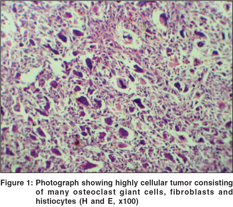
Indian Journal of Dermatology, Venereology and Leprology, Vol. 72, No. 2, March-April, 2006, pp. 145-146 Case Report Giant cell tumor of skin Y Sunil Kumar, Raghupathi AR, Chidananda Department of Pathology, Government Medical College, Mysore, Karnataka Code Number: dv06044 Abstract Giant cell tumor of the skin is a rare entity showing gross and histological features similar to those of giant cell tumor of the bone. We report a case of malignant giant cell tumor of the thigh in a 55-year-old man. Histological features showed a biphasic population of mononuclear cells admixed with osteoclast-like giant cells. The nuclei of the giant cells were similar to those of the mononuclear cells. This tumor should be distinguished from a variety of cutaneous neoplasms that contain multinucleated giant cells.Keywords: Giant cell tumor, Malignant, Skin Introduction Giant cell tumor of the skin is a rare entity showing similar gross and histological features of giant cell tumor of the bone.[1] Histologically, these lesions show round to spindle shaped cells intimately admixed with uniformly scattered osteoclast-like multinucleate giant cells.[2] Superficial tumors involving either the subcutis or the fascia have a much better prognosis than deeply situated ones. We report a patient with this rare neoplasm.Case Report We were sent a specimen for histopathological examination by a physician from a remote place who had done an excisional biopsy of a swelling. The patient was a 55-year-old man who had an asymptomatic swelling on the upper right thigh since 6 months. The swelling measured 5 x 3.5 x 2 cm and was mobile, nontender and adherent to the skin. There was no regional lymphadenopathy. An X-ray showed no bony involvement. Grossly, the specimen consisted of a skin-covered irregular mass measuring 4.5 x 3 x 1.5 cm. Histologically, there was a well-circumscribed multinodular area involving the dermis and consisting of fibroblasts, histiocytes and osteoclast-type giant cells [Figure - 1]. The mononuclear cells showed pleomorphism and increased mitotic activity. Immunohistochemistry studies revealed osteoclast cells positive for CD68 and negative for EMA, vimentin and cytokeratin markers. A diagnosis of malignant giant cell tumor of the skin was made; however, the patient did not follow up later. Discussion Primary giant cell tumor of soft tissue, also known as giant cell tumor of low malignant potential, is a rare soft tissue tumor located in both the superficial and deep soft tissues.[2] The lesion usually affects patients aged between 68 and 78 years (median 73 years) with a male to female ratio of 3:2.[1],[3] It commonly involves the extremities, head and neck. Its gross and histologic features are similar to the giant cell tumor of the bone. Most tumors are well circumscribed, unencapsulated and multinodular with a mixture of histiocytes, fibroblasts and osteoclast-like giant cells. The fibroblasts show some tendency to aggregate at the periphery of the nodule. The fibroblasts and histiocytes display pleomorphism with increased mitotic activity and often contain ingested lipid and hemosiderin. Osteoclasts arise due to fusion or amitotic division of these mononuclear precursors. The giant cells have voluminous eosinophilic cytoplasm with numerous small uniform nuclei. Phagocytic vacuoles and asteroid bodies are occasionally present.[2] In approximately 50% of cases, focal osteoid or mature bone is present, usually located at the periphery of the tumor. It appears to be produced by neoplastic cells. The histogenesis is unclear. This tumor was earlier included as one of the histological types of malignant fibrous histiocytoma, but this view is no longer favored. The differential diagnosis includes benign fibrous histiocytoma, atypical fibroxanthoma, giant cell tumor of bone with soft tissue extension, leiomyosarcoma with osteoclast giant cells and extraskeletal osteosarcoma. Benign fibrous histiocytoma with many osteoclast giant cells can be differentiated by the presence of hyperplastic epidermis, hyperpigmentation of the basal layer and elongation of rete ridges, separated by clear (Grenz) zone from the spindle cell tumor in the dermis.[4] Atypical fibroxanthoma shows pleomorphic histiocyte-like cells and atypical giant cells, often with bizarre nuclei and numerous mitotic figures.[2],[4] Giant cell tumor of bone with soft tissue extension shows radiologically, an osteolytic lesion in the epiphysis and the presence of a rim of ossification at the edge of the tumor.[5] Leiomyosarcoma with osteoclast giant cells shows brightly eosinophilic spindle cells with vesicular, cigar shaped nuclei.[2] Extraskeletal osteosarcoma can be differentiated by the presence of neoplastic bone or osteoid.[6] Superficial tumors involving the subcutis or fascia have a much better prognosis than deeply situated ones; 75% of superficial tumors recur and only 25% metastasize, while about 50% of deep tumors recur and about 50% metastasize.[2] Recently, two cases of malignant giant cell tumor in the dermis extending into the subcutaneous tissue were reported.[7] References
Copyright 2006 - Indian Journal of Dermatology, Venereology and Leprology The following images related to this document are available:Photo images[dv06044f1.jpg] |
| |||||||||
