
|
Indian Journal of Dermatology, Venereology and Leprology
Medknow Publications on behalf of The Indian Association of Dermatologists, Venereologists and Leprologists (IADVL)
ISSN: 0378-6323 EISSN: 0973-3922
Vol. 72, Num. 2, 2006, pp. 161-164
|
Indian Journal of Dermatology, Venereology and Leprology, Vol. 72, No. 2, March-April, 2006, pp. 161-164
Resident's Page
Scaly signs in dermatology
Kangle Satyajeet, Amladi Sangeeta, Sawant S
Department of Dermatology, B. Y. L. Nair Chartiable Hospital, Mumbai
Correspondence Address:Dr. Satyajeet D. K., Department of Dermatology, B. Y. L. Nair Charitable Hospital, Mumbai Central, Mumbai 400 008, Maharashtra, India. E-mail: satyajeetdk@yahoo.co.in
Code Number: dv06054
Goethe: What is most difficult of all? It is what appears most simple: To see with your eyes what lies in front of your eyes.[1]
Unlike other specialties such as internal medicine or surgery, dermatologists have the advantage of direct examination of the lesions that occur on the surface of skin. This visibility of skin lesions often enables the physician to make an instant or spot diagnosis. Scaling is an important secondary lesion in dermatology. The word scale is derived from skal ( Old English - Scealu; Old French - escale; or French - ιcaille - shell; Latin -
Squama - a scale or plate-like structure).[2]
Gentle scratching and rubbing alters visibility of scaling. Scratching
scale in psoriasis makes the scale appear more silvery in color by
introducing air-keratin interfaces.
On grattage, characteristic coherence of the scales can be seen as
if one scratches a wax candle - signe de la tache de bougie.
In non-scaly lesions, indentation by a fingernail leaves an opaque
mark resembling that made by scratching a tallow wax candle.
Signs in scaling
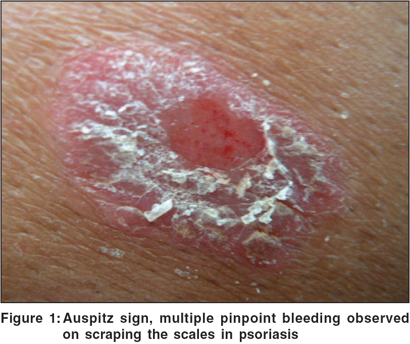
Auspitz sign
Heinrich Auspitz (1835-1886) was the early star among Ferdinand Ritter
von Hebra′s pupils. But this sign was already described by number
of authors including Hebra, Robert Willan and Daniel Turner. However,
the Auspitz phenomenon is eponymously linked to him because his extraordinary
treatise on general pathology and therapeutics of the skin was translated
into English in 1885 and thereby constituted an early harbinger of
central European dermatopathology.[3]
When the scales are completely scraped off, the stratum mucosum (basement membrane) is exposed and is seen as a moist red surface (membrane of Bulkeley) through which dilated capillaries at the tip of elongated dermal papillae are torn, leading to multiple bleeding points [Figure - 1]. This is a characteristic feature of psoriasis and is known as Auspitz sign. It is attributed to parakeratosis, suprapapillary thinning of the stratum malphighii, elongation of dermal papillae and dilatation and tortuosity of the papillary capillaries.
However, Auspitz sign is not sensitive or specific for psoriasis.[4] Not
sensitive, because in one study, out of 234 patients it was seen in 41
patients of psoriasis. Also it is not seen in inverse psoriasis; pustular,
erythrodermic psoriasis; guttate psoriasis. Not specific because it is
also seen in nonpsoriatic scaling disorders, including Darier′s
disease and actinic keratosis.
Carpet tack sign (cat′s tongue sign, tin tack sign)[5]
In DLE, characteristic lesions are well-defined erythematous plaques
with partially adherent scales entering a patulous follicle. When the
scale is removed, its undersurface shows horny plugs that had occupied
follicles. This is called the carpet tack or tintack sign.
However, carpet tack sign is not diagnostic of DLE. It is also seen
in seborrheic dermatitis and pemphigus foliaceous. But in DLE, on
removal of scale, bleeding may be seen due to adherent scales unlike
in pemphigus
foliaceous/seborrheic dermatitis, where the scales are loose.
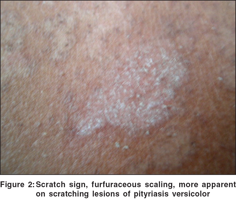
Scratch sign (coup d′ongle sign, besnier′s sign, stroke
of the nail)
Pityriasis versicolor is characterized by asymptomatic hypopigmented
or hyperpigmented macules and patches and produces fine scales
(branny/furfuraceous). Often the scale is not visible. An important
diagnostic clue may
be the loosing of barely perceptible scale with a fingernail, which
is
called
as the scratch sign [Figure - 2]. This sign may be negative if
patient has taken recent bath or in case of treated lesion, in which
case,
only hypopigmentation persists.
Scaling is the common finding in disorders characterized by scaling
(squamous) papules, plaques and patches, which are often termed
papulosquamous. Scale is usually white or light tan and flakes
off rather easily.
This should be distinguished from crust, which is dried serum
and debris
on
the skin surface. The distinction between a scale and a crust
is important because the differential diagnosis is entirely different
for the two.[6]
Types of scale
Collarette scale
Describes the fine, peripherally attached and centrally detached scale at the edge of salmon-colored patch/plaque. Examples: Pityriasis rosea, subsiding lesions of furuncle, miliaria, erythema nodosum, etc.
Furfuraceous scale (Latin furfur - bran)
Describes fine and loose scales that are not conspicuous and made visible
by scratching (scratch sign). Example: Pityriasis versicolor.
Ichthyosiform scale
Describes large, polygonal scales - as in fish scales. Example: Ichthyosis
vulgaris.
Micaceous scale (Silvery)
Describes a silvery, white, parakeratotic, lamellated scale. Silvery
white appearance is due to reflection of light at the air-keratin interface
between the layers of scale. Example: Psoriasis vulgaris.
Greasy scale
Describes loose, moist, yellow-brown oily scaling, especially perifollicular,
on seborrheic areas. Example: Seborrheic dermatitis. N.B.: In Darier′s
disease greasy, dirty, warty excrescences are distinctly papular with crusts;
besides, associated nail changes, palmar pits and cobble stoning can be
seen.
Trailing scale
Describes annular erythema with advancing flat or elevated border and trailing
scale at the inner border with central area flattening and fading. Lesions
occur on trunk and especially, buttocks, inner thighs. Example: Erythema
annularis centrifugum.
Wafer like scale
Thin adherent mica-like scale attached at the center of a lichenoid firm
reddish brown papule and free at the periphery. Example: Pityriasis lichenoides
chronica. In clear cell acanthoma, wafer-like scale is seen adherent at
the periphery, which leaves a moist or bleeding surface when removed.
Double-edged scale
Describes erythematous, exfoliating or scaly, annular or polycyclic, flat
patch with an incomplete advancing double edge of peeling scale. Example:
Ichthyosis linearis circumflexa (ILC). [Netherton syndrome=ILC + hair abnormality + atopic
diathesis]
Cornflake sign/scale
Sometimes used for scale crust of pemphigus foliaceous. Cornflake
sign seen in Flegel′s disease is characterized by 2-3 mm keratotic
scaly papules with discrete irregular margins. The scale separates from
many lesions, leaving a non-exudative red base.
Scales in erythoderma
Depending on the stage of erythroderma - acute or chronic - scales can
be large plate-like sheets in acute stage or fine and bran-like in chronic
stage.
Hystrix-like scale
Porcupine spine describes muddy brown or gray color scaling over verrucous
lesion, either generalized or nevoid, commonly affecting extensor aspects
of the limbs, truncal areas to variable degrees. Example: Ichthyosis hystrix.
Mauserung desquamation
Describes circumscribed patchy scaling with focal desquamation or moulting
of scale that Siemens called mauserung, seen at the flexures and acral
sites, especially the dorsal hands and feet. Example: Ichthyosis bullosa
of Siemens (mild variant of BIE).
Carapace-like scale
Described as white or gray, small, flaky or branny and semi-adherent scale
with turned-up edges, seen on extensor surfaces of the arms and lower legs
and characteristically spares the flexural creases. Sometimes fine scales
have ′pasted-on appearance.′Example: Ichthyosis vulgaris.
Coat of armor
The affected infant is encased in a rigid, taut, yellow-brown adherent
skin, a hyperkeratotic coat of armor covering the whole body. Example:
Harlequin ichthyosis.
Plate-like scale (Armor plate)
Described as large, polygonal, thick, rigid, dark brown or gray firmly
adherent scales, which appear to be arranged in a mosaic pattern but
tend to be largest over the lower extremities, where it may give an
appearance of dry riverbed. Example: Lamellar ichthyosis.
Corrugated/Ridged scale
In bullous ichthyosiform erythroderma, as erythroderma and blistering
tendencies diminish, the characteristic gray waxy scale progresses.
Yellow-brown, waxy, ridged or corrugated scale builds up in skin
creases, namely, anterior
neck, flexures, abdominal wall, infra-gluteal folds and scalp.
Latent desquamation
Scale formation can sometimes be observed only after scratching the
lesion - may be found in the early stages of pityriasis rosea as
well as in
pityriasis versicolor, parapsoriasis and psoriasis.
Oyster-like scale
Large heaped-up scale accumulation in psoriasis is described as
ostraceous or oyster-like scale.
Sandpaper-like
In actinic keratosis, the firmly adherent, dry, rough and often
yellow or brown colored scales have a gritty feel like sandpaper
and the
scales are better appreciated by skin palpation.
Accurate clinical diagnosis is based on vigilant observation
for morphology and pattern of lesions and elicitation of
clinical signs.
In the coming
years, more and more such clinical signs are likely to improve
diagnostic acumen of dermatologists.
References
| 1. | Stewart MI, Bernhard JD, Cropley TG, Fitzpatrick TB. The structure of skin lesion and fundamentals of diagnosis. In: Freedberg IM, Eisen AZ, Wolff K, Austen KF, Goldsmith LA, Katz SI, editors. Fitzpatrick's Dermatology in general medicine. 6th ed. McGraw-Hill: New York; 2003. p. 11-30. Back to cited text no. 1 |
| 2. | Scale (definition). www.skincareguide.ca/glossary/s/scale.html Last accessed on 23rd March 2006. Back to cited text no. 2 |
| 3. | Holubar K . Papillary tip bleeding or the Auspitz phenomenon: A hero wrongly credited and a misnomer resolved. J Am Acad Dermatol 2003;48:263-4. Back to cited text no. 3 |
| 4. | Bernhard JD. Auspitz sign is not sensitive or specific for psoriasis. J Am Acad Dermatol 1990;22:1079-81. Back to cited text no. 4 [PUBMED] |
| 5. | Goodfield MJ, Jones SK, Veale DJ. The connective tissue diseases. In: Burns T, Breathnach S, Cox N, Griffiths C, editors. Rook's Textbook of dermatology. 7th ed. Blackwell Science: Oxford; 2004. p. 56-9. Back to cited text no. 5 |
| 6. | Lookingbill DP, Marks JG. Principles of clinical diagnosis. In: Moschella SL, Hurley HJ, editors. Moschella's Dermatology. 3rd ed. WB Saunders Co: Philadelphia; 1992. p. 165-239. Back to cited text no. 6 |
Copyright 2006 - Indian Journal of Dermatology, Venereology and Leprology
The following images related to this document are available:
Photo images
[dv06054f2.jpg]
[dv06054f1.jpg]
| 