 |
| About Bioline | All Journals | Testimonials | Membership | News |
|
||||||
|
||||||
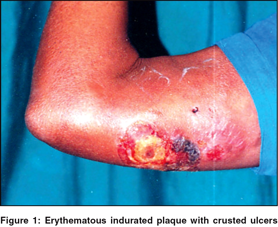
Indian Journal of Dermatology, Venereology and Leprology, Vol. 72, No. 3, May-June, 2006, pp. 215-217 Case Report CD-3 positive extranodal T-cell lymphoma of nasal type with skin involvement Reddy RaghunathaR, Singh Gurcharan, Prathima KJ, Harendra Kumar ML Departments of Dermatology, Sri Devaraj Urs Medical College, Tamaka, Kolar, Karnataka Code Number: dv06071 Abstract A 40-year-old previously healthy lady presented with nasal obstruction and localized plaques over the right arm. She developed complete nasal obstruction due to a mass in the right nasal cavity and skin lesions that ulcerated to present as ecthyma gangrenosum like lesions. Patient's condition deteriorated fast and she developed icterus with fatal outcome within 4 weeks of developing skin lesions. Nasal and skin biopsy revealed angiocentric T-cell lymphoma, which on immuno-phenotyping revealed CD-3 positive; and CD-20, CD-30, ALK and EMA negativity. She was seronegative for HIV. Final diagnosis of CD-3 positive extranodal T-cell lymphoma of nasal type was made. Extranodal T-cell lymphomas are very aggressive NHLs with poor prognosis. Prognosis depends on histology, stage of the disease and sites of involvement. NK/T cell lymphoma of nasal type is common with EBV association. Skin involvement is rare and is also an indicator of poor prognosis.Keywords: Angiocentric lymphoma, CD-3 positive, Extranodal T-cell lymphoma Introduction Non-Hodgkin's lymphomas (NHLs) presenting as skin lesions are more commonly of B-cell phenotype, followed by T-cell phenotype and mycosis fungoides.[1] B-cell type of lymphomas in the category of extranodal type in the cervicofacial region are in greater majority than the T-cell type and most of them are associated with EBV infection and are more common in renal allograft patients.[2] Extranodal involvement - especially of skin, irrespective of the extent of involvement - is an important poor prognostic factor.[3] Though skin involvement is rare, it has been reported and a high index of suspicion is required for early diagnosis. We report a case of CD-3 positive extranodal T-cell lymphoma of nasal type with skin involvement in a 40-year-old previously healthy lady, with rapid progression resulting in a fatal outcome. Case report A 40-year-old lady presented with history of nasal obstruction, which gradually increased over a period of 4 months. She developed localized swelling and skin lesions over right arm with pain. Later she developed fever, icterus and the nasal obstruction worsened. She had no history of significant illness or surgery in the past. General physical examination was normal except for the presence of icterus and there was no regional lymphadenopathy. On examination, localized indurated, ill-defined plaques were seen over an erythematous base over the posteromedial aspect of the right arm, measuring approximately 8 cm x 10 cm in diameter. Subsequently, tenderness, necrosis, crusting and ulceration developed over the plaques [Figure - 1]. ENT examination revealed reddish mass in the right nasal cavity, adherent to the lateral wall and bleeding on touch. The same mass was seen in the nasopharynx on posterior rhinoscopy and a clinical diagnosis of nasopharyngeal carcinoma was made. Following were the results on investigations: Hb - 9 gm%; total count - 5000 cells/mm 3; differential count: N - 82%, L - 15%, E - 02%, M - 01%; ESR - 80 mm/Hr; Blood culture - no yield; Mantoux test - negative; ELISA for HIV - negative; HbsAg - negative; serum bilirubin: total - 10.9 gm%, direct - 6.2 gm%. Ultrasound examination revealed mild hepatosplenomegaly with cholecystitis. CT scan revealed a mass in the nasopharynx extending to involve the skull base, right maxillary antrum and right nasal cavity. Skin biopsy and endoscopic biopsy of the nasal mass showed groups and sheets of round to oval tumor cells with a vesicular nucleus and prominent nucleoli, admixed with mononuclear inflammatory cells. The tumor cells were infiltrating the vessel wall with destruction of the wall and areas of necrosis. A histopathological diagnosis of angiocentric lymphoma was made based on the above findings. Immunophenotyping of the same specimen revealed CD-3 positive; and CD-20, CD-30, ALK and EMA negativity; and a final diagnosis of extranodal CD-3 positive T-cell lymphoma of nasal type with skin involvement was made. Three weeks after developing skin lesions and after debridement, she lapsed into semiconsciousness, later coma and died within 4 weeks after developing skin lesions. Discussion Based on the above findings, this case was classified as extranodal peripheral T-cell lymphoma of nasal type, but EBV association could not be established and natural killer cell phenotype was not confirmed due to technical difficulties. Extranodal NK/T-cell lymphoma of nasal type has been defined in the recently proposed WHO classification of hemopoietic and lymphoid neoplasms. They are almost universally associated with EBV infection.[2] Most common localization of NK/T-cell lymphoma is in the head and neck region, especially the nasal cavity and it can also occur in other sites like skin and gonads.[2],[4] Histopathologically, they can be intermediate or high-grade malignancies and should be differentiated from squamous cell carcinomas due to their clinical similarity. Morphologically, they were previously termed as angiocentric lymphomas (Revised European-American Lymphoma classification).[5] Clinically, they are very aggressive neoplasms with poor prognosis.[2],[6] This case resembles a recently described clinicopathological syndrome of fulminant T-cell lymphoproliferative disorder following acute/chronic EBV infection, characterized by fever, hepatosplenomegaly, pancytopenia, erythrophagocytosis and liver failure.[7] NHLs presenting as skin lesions are more commonly of B-cell phenotype.[1] Presence of extranodal disease at the time of presentation is the most important clinical variable and portends a poor prognosis.[3] Treatment consists of radiation and/or chemotherapy. Prognosis depends on histology, stage and site of lesion. Newer treatment strategies may lead to improved survival for patients with head and neck NHLs.[8] This case is being reported due to its rarity, aggressive course, rapid fatality, skin and, probably, hepatic system involvement. A high degree of clinical suspicion and complete workup in a short span is required to diagnose this entity. References
Copyright 2006 - Indian Journal of Dermatology, Venereology and Leprology The following images related to this document are available:Photo images[dv06071f1.jpg] |
| |||||||||
