 |
| About Bioline | All Journals | Testimonials | Membership | News |
|
||||||
|
||||||
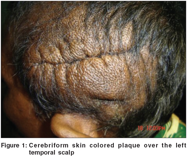
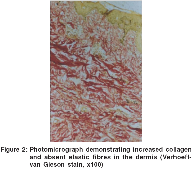
Indian Journal of Dermatology, Venereology and Leprology, Vol. 72, No. 4, July-August, 2006, pp. 309-311 Letter To Editor Isolated scalp collagenoma mimicking cutis verticis gyrata Laxmisha Chandrashekar, Thappa DevinderMohan, Jayanthi S Department of Dermatology and STD, Jawaharlal Institute of Postgraduate Medical Education and Research (JIPMER), Pondicherry Code Number: dv06105 Sir, Cutis verticis gyrata (CVG) is a descriptive term for a condition of the scalp, in which deep furrows and convolutions are seen, that resembles the outer surface of the cerebrum. The etiology is diverse, since different collections of cell types may be responsible for outward convoluted appearance, and range from inflammatory or hamartomatous infiltrations to neoplastic proliferations.[1] Collagenoma or connective tissue nevi of the collagen type are hamartomatous lesions, consisting of proliferation of normal collagen tissue. They can be hereditary or sporadic. The lesions consist of slightly elevated nodules that may be grouped or disseminated. Collagenomas located in the plantar or palmar surface with a cerebriform appearance are rare, and have been reported in Proteus syndrome.[2],[3] Herewith, isolated scalp collagenoma mimicking cutis verticis gyrata is being reported for its rarity and unique localization. A 35-year-old female presented with asymptomatic, asymmetrically located, solitary, cerebriform skin colored plaque of 18´12 cm over the left temporal scalp since birth [Figure - 1]. The plaque had been gradually increasing in size. There was no other skin, mucosal, soft tissue, or bone abnormalities. There was no family history of similar disorder. A representative skin biopsy specimen (scalp) revealed a normal epidermis, and an increased amount of collagen fibres within the papillary and reticular dermis. Masson trichrome stain showed a significant increase of thick collagen bundles, with some of them in vertical bundles. Verhoeff- van Gieson stain showed absent elastic fibers [Figure - 2]. Skeletal survey did not demonstrate any evidence of osteopoikilosis. Routine investigations were normal. Systemic examination did not reveal any features suggestive of neurocutaneous syndromes or endocrine disorders. Thus, a diagnosis of isolated collagenoma of the scalp was made. Connective tissue nevi of the hereditary type include dermatofibrosis lenticularis disseminata in the Buschke Ollendorff syndrome, familial cutaneous collagenoma, and shagreen patches seen in tuberous sclerosis. Connective tissue nevi of acquired type have been classified as eruptive collagenomas, isolated collagenomas, or isolated elastomas, depending on the number of lesions and the predominant dermal fibers present.[4] Familial cutaneous collagenoma is characterized by a symmetrical eruption of collagen-type nodules and plaques occurring predominantly over the upper back in adolescence. Shagreen patches are present in tuberous sclerosis, in association with the classic skin findings of adenoma sebaceum, periungual fibromas, and ash-leaf macules.[4] Plantar collagenomas with cerebriform appearance have been described as one of the major skin findings of Proteus syndrome. However, three cases of isolated plantar collagenoma without associated clinical abnormalities have been reported.[4] In one case of isolated collagenoma, Uitto et al[5] showed that the increased collagen is of the adult type (Type I) and that a local reduction of collagenase might be the cause of the excess collagen. In contrast to cutis verticis gyrata (CVG) with a diffuse involvement of scalp, isolated collagenoma presents as a localized abnormality. Biopsies from CVG usually show a thickened dermis with possible sebaceous hyperplasia, with or without collagen excess, whereas isolated collagenoma only shows excess of collagen without sebaceous hyperplasia. To the best of our knowledge, we could not find such a report of isolated collagenoma of the scalp presenting as cutis verticis gyrata, in the published literature. In summary, collagenoma may be a marker for internal disease such as tuberous sclerosis, may occur in familial or eruptive patterns, or may be present in isolation, as in our case. As such, isolated cerebriform scalp collagenoma mimicking cutis verticis gyrata is unique, and may be considered as secondary cause of cutis verticis gyrata. References
Copyright 2006 - Indian Journal of Dermatology, Venereology and Leprology The following images related to this document are available:Photo images[dv06105f1.jpg] [dv06105f2.jpg] |
| |||||||||
