 |
| About Bioline | All Journals | Testimonials | Membership | News |
|
||||||
|
||||||
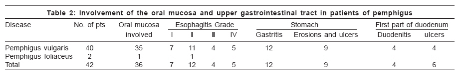
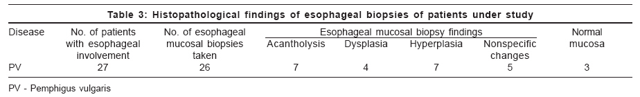
Indian Journal of Dermatology, Venereology and Leprology, Vol. 72, No. 6, November-December, 2006, pp. 421-424 Original Article Study of upper gastrointestinal tract involvement in pemphigus by esophago-gastro-duodenoscopy Rao PNarasimha, Samarth Aruna, Aurangabadkar SanjeevJ, Pratap Bajrang, Lakshmi TSS Department of Dermatology, Osmania Medical College, Hyderabad Code Number: dv0647 Abstract Introduction: Involvement of upper gastrointestinal tract in pemphigus vulgaris is not uncommon.Aim: To study the involvement of upper gastrointestinal tract (UGIT) with the help of esophago-gastro-duodenoscopy (EGD) in patients of vesiculobullous dermatoses with emphasis on pemphigus vulgaris. Methods: Forty-two patients (M-22, F-20) with vesiculobullous dermatoses, diagnosed on the basis of clinical features and skin histopathology as pemphigus vulgaris (PV)-40 patients and pemphigus foliaceus (PF)-2 patients were included in the study. The EGD was performed and mucosa of the esophagus, stomach and first part of the duodenum were examined. Mucosal biopsies were taken from the lower esophagus in 26 patients of PV and studied after H and E staining. Results: On EGD, esophageal involvement was seen in 67% patients of PV (27/40). Of these, Grade I esophagitis was observed in seven, Grade II in 11, Grade III in four and Grade IV involvement was seen in five patients of PV. Three PV patients had associated esophageal candidiasis. Involvement of esophageal mucosa was also observed in one out of two patients of PF. Gastric mucosa was involved in 52% and duodenal mucosa in 20% of PV patients. Acantholysis was observed in seven out of 26 (27%) esophageal biopsies of PV patients. Two patients of PV vomited a tube-like structure, indicative of 'esophagitis dissecans superficialis'. The involvement of the gastric mucosa in patients with history of oral corticosteroid intake (60%) was compared to the group without history of oral corticosteroids (30%). Conclusion: Among PV patients under study, significant involvement of oral (87%), esophageal (67%), gastric (52%) and duodenal mucosa (20%) was observed. Keywords: Esophago-gastro-duodenoscopy, Esophagus, Gastric mucosa, Pemphigus vulgaris, Vesiculobullous disorders Introduction Vesiculobullous dermatoses, particularly the pemphigus group of disorders, are important for the morbidity associated with the involvement of skin and mucous membranes. In the diseases where they are involved, mucosal involvement is not limited only to the oral cavity but extends to the esophagus as well. This upper gastrointestinal tract (UGIT) involvement adds to the morbidity and complications of the disease. Esophago-gastro-duodenoscopy (EGD) or upper gastrointestinal endoscopy is one of the most commonly performed endoscopic procedures. In the present study, we examined patients of pemphigus for the involvement of the UGIT with the help of this procedure and in some patients mucosal biopsies from esophagus were studied. Methods A total of 42 patients (22 males, 20 females) presenting with pemphigus to the Department of Dermatology, Osmania General Hospital, Hyderabad, India, between January 1995 and December 1999 were included in the study. Both untreated and partially treated patients of all age groups were included in the study after fully informed written consent was taken. Detailed clinical examination was performed and recorded. Skin biopsies were taken from active skin lesions in all patients to confirm clinical diagnosis. Direct or indirect immunofluoroscence was not performed due to lack of facilities. All the patients were subjected to EGD at the Department of Gastroenterology, Osmania General Hospital, Hyderabad. The severity of involvement of the esophageal mucosa was graded on a scale of I to IV based on the endoscopic findings [Table - 1]. Similarly, the macroscopic findings under EGD of the gastric and duodenal mucosa were recorded. Biopsies from the esophageal mucosa were taken from active lesions involving the lower esophagus in 26 patients.Results Out of the 42 patients, 22 were male and 20, female. The age distribution of the patients was as follows: five patients were in the 10-20 years age group, 23 were in the 20-45 years age group and 14 were aged over 45 years. The clinical diagnosis was PV in 40 patients and PF in two patients. The duration of the disease in these patients ranged from one month to four years. On clinical examination, the oral mucosa was involved in 35 out of 40 patients of PV patients and in one out of two patients of PF. It was predominantly in the form of erosions and ulcers of the buccal mucous membrane; in 12 patients, the lips were also involved. The majority of the patients with oral ulcers complained of burning pain, dysphagia and odynophagia. One patient of PV gave a history of hematemesis. The results of EGD are given in [Table - 2]. On EGD, esophageal involvemnt was observed in 27 out of 40 PV patients and one out of 2 PF patients. Three patients with Grade IV esophagitis of these patients also had associated esophageal candidiasis. On EGD, gastric mucosal involvement was observed in 21 patients and duodenal mucosal involvement in eight [Table - 2]. The types of lesions involving the antrum and body of the stomach were ulcers, erosions and severe diffuse gastritis. In the duodenum, duodenitis, small ulcers and deformed cap were the types of involvement. None of the PF patients showed gastric or duodenal involvement. Esophageal mucosal biopsies were taken in 26 patients with esophageal involvement [Table - 3]. Twenty eight of the 42 patients had taken oral corticosteroids for up to six months for their disease before presenting to us and undergoing EGD. Gastric mucosal involvement was commoner in patients taking oral corticosteroids than in those who had not, while esophageal involvement was equally common in both these groups. Another significant observation was the vomiting of a tube-like structure, indicative of esophagitis dissecans superficialis, by two patients of PV. Discussion An assessment of UGIT involvement in pemphigus may help determine the extent and severity of the disease and may guide the approach to therapy.[1] We observed esophageal involvement in 27 out of 40 patients of PV (67%). In a study from India, 18 out of 25 (72%) PV patients had esophageal involvement.[2] All PV sera have anti-Dsg3 antibodies and over 50% have anti-Dsg1 antibodies. Antibodies against both Dsg3 and Dsg1 are required to produce significant blistering in the skin.[3] Pemphigus vulgaris entirely limited to the mucous membranes is seen in those with no reactivity to Dsg1.[4] In a majority of the patients, painful oral mucous membrane erosions are the presenting sign of PV.[5] In our study, 87% of patients of PV presented with oral lesions. About half of the patients of PV have esophageal involvement but this is usually asymptomatic.[1] The whole or segments of the esophageal mucosa can be involved.[6] Patients may complain of odynophagia, dysphagia and occasionally gastrointestinal bleeding. In our study, 36 patients with oral mucosal ulcers and erosions (including 27 with esophageal involvement) presented with varying grades of dysphagia and odynophagia. One patient also gave a history of hematemesis. Esophageal involvement may become evident in the absence of cutaneous manifestations when the latter are in remission or during treatment.[7],[8] Very rarely, bullous diseases may affect the esophagus in such a manner that there is sloughing of the entire mucous membrane, leading to the formation of a mucosal cast (′esophagitis dissecans superficialis′), which may be vomited.[9] Such a condition was observed in two of our patients; one of them who vomited this cast did so on the second day after his EGD, which probably precipitated the cast′s dislodgement. Acantholysis was observed only in seven out of 26 esophageal biopsies (27%) in our study. In similar studies, acantholysis was observed in 36% and 50% of PV patients.[1],[10] We could not perform direct immunofluorescence (DIF) on esophageal biopsies although studies have shown that DIF is positive in all PV patients biopsied from any part of the esophageal mucosa, demonstrating that immune deposition occurs along the entire length of the esophagus irrespective of the degree of clinical involvement.[10] A finding either to unreported was the involvement of the gastric (52%) and duodenal (20%) mucosa on EGD in our patients of PV. Stress ulcerations of gasric mucosa are known to occur in very ill patients due to the loss of barrier protection during critical illness. We did not perform any histological or immunological study of gastric or duodenal mucosal lesions. We could only presume that these lesions if not due to the disease, could be due to the effects of oral corticosteroids as many patients gave history of their intake. It has been observed that lack of correlation between the patients′ subjective complaints and endoscopic findings indicates the importance of endoscopy in assessing gastroduodenal lesions in patients on corticosteroid therapy.[11] In our study, 28 out of 42 patients gave a history of taking oral steroids for varying periods before undergoing EGD. Gastric mucosal involvement was higher in this group of patients (60%) than in those who had not taken oral steroids (28%), indicating that steroid intake played a role.
Three patients in the present study demonstrated candidiasis of esophagus on EGD. Prolonged use of corticosteroids, including topical preparations, have reportedly led to oropharyngeal and esophageal candidiasis in otherwise healthy individuals.[14] Hypochlorhydria due to acid-suppressive medications, which are usually given as adjuvants along with corticosteroids, predisposes to candidial esophagitis by removing the cleansing effect of normal periodic acid reflux events.[15] In the present study, we have observed a high proportion of oral and esophageal mucosal involvement in PV patients, apart from involvement of the stomach and duodenum. Study of the esophagus by EGD in PV and other vesiculobullous disorders is not only useful in assessing the extent of the involvement of UGIT but also in evaluating the efficacy of the treatment instituted. The lesions in the stomach and duodenum observed in patients of PV in our study cannot be attributed to the disease as the histopathology or immunopathology of these lesions was not performed. However, reports of the involvement of the colon in a patient of pemphigus[13] entails that further studies are required to determine the extent and nature of involvement of gastro intestinal mucosa in these disorders. References
Copyright 2006 - Indian Journal of Dermatology, Venereology and Leprology The following images related to this document are available:Photo images[dv06147t2.jpg] [dv06147t1.jpg] [dv06147t3.jpg] |
| |||||||||
