 |
| About Bioline | All Journals | Testimonials | Membership | News |
|
||||||
|
||||||
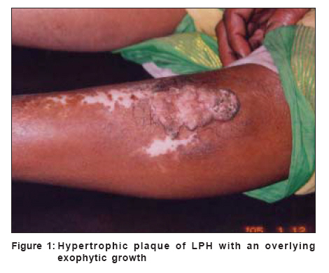
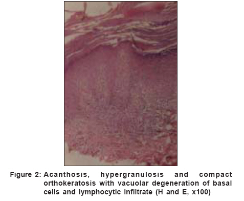
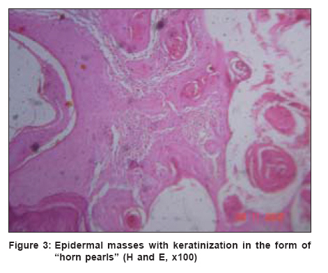
Indian Journal of Dermatology, Venereology and Leprology, Vol. 72, No. 6, November-December, 2006, pp. 470 Net letter Malignant transformation of hypertrophic lichen planus Sengupta Sujata, Das JayantaKumar, Gangopadhyay Asok Department of Dermatology, RKM Seva Pratisthan and Vivekananda Institute of Medical Sciences, Kolkata Code Number: dv06164 Sir, Lichen planus (LP) is a common papulosquamous disorder involving the skin, nails and mucosae.[1] A variant of LP is the hypertrophic or warty type better known as lichen planus hypertrophicus (LPH). Although it is a benign disorder, malignant changes may happen and till 1997, only 30 such cases have been reported.[2] A middle-aged woman who developed squamous cell carcinoma (SCC) over a preexisting lesion of LPH is described here for the rarity of the condition. A 58-year-old housewife presented with a warty growth over the anterior aspect of her left leg since the last 10 years. The lesion had started as a small red itchy plaque that gradually enlarged to attain its present size. Over the last six months, a small, hard growth had appeared at its upper end that enlarged insidiously and sometimes bled a little on manipulation. There was no history of trauma or any application of counter-irritant substances at the local site. She was otherwise of normal health and a similar disease was not known in the family. On examination, a pigmented hypertrophic plaque, 7 ´ 4 cm, with a well-defined raised border and an irregular surface was seen on the anterior aspect of the left leg, a little below the knee joint. An exophytic growth, 1.5 ´ 2 cm, was seen overlying the upper end of the plaque [Figure - 1]. It had elevated, noneverted margins with a crater-like depression in the center. The growth was fixed to the underlying skin and soft tissue. There was evidence of healing in a few areas that showed depigmentation with mild atrophy. The mucous membranes, hair and nails were unaffected. Regional lymph nodes were not palpable and the rest of the systemic examination was normal. After routine hematological and biochemical investigations, all of which turned out to be normal, a chest skiagram and abdominal ultrasound was done. Evidence of lymph node enlargement or organ metastasis was not found. Biopsy specimens were obtained from the edge of the plaque and from the overlying tumor. The first one showed marked irregular acanthosis, hypergranulosis and vacuolar degeneration of the basal cells at the base of rete ridges with a sparse lymphocytic infiltrate. These findings were typical of LPH [Figure - 2]. The second specimen showed epidermal proliferation made up of mature keratinocytes with horn pearls and scattered atypical mitotic figures [Figure - 3]. The features were of a well-differentiated SCC. The longstanding plaque of LPH had undergone malignant transformation into a SCC. The tumor was treated with complete excision and closed with an autograft taken from the thigh. Although the incidence of cancer in oral LP is about 1.3%, neoplastic transformation in cutaneous LP is very rare.[3] The underlying mechanism of this malignant conversion is not exactly known but speculatively, chronic inflammatory processes show an overdrive of growth factors that constantly stimulate epithelial cell proliferation into neoplastic conditions.[3] There are reports of a few such cases from India and the patients had multiple lesions of LP, one or two of which underwent malignant change.[4] In our case, in spite of the long duration of the disease, there was a solitary lesion. Histopathology in such lesions shows marked hyperkeratosis, acanthosis, papillomatosis and hypergranulosis.[5] Sometimes, such cases may mimic SCC clinically but histopathology reveals LPH with pseudo-carcinomatous hyperplasia without any evidence of malignancy. However, these cases require a regular and vigilant follow-up. Our patient had a well-differentiated SCC with a size less than 2 cm and absence of overlying ulceration, which are indicators of a good prognosis and she has been doing well with the treatment given. Hence, careful vigilance of a longstanding LPH is necessary to allow early detection of a developing SCC. References
Copyright 2006 - Indian Journal of Dermatology, Venereology and Leprology The following images related to this document are available:Photo images[dv06164f3.jpg] [dv06164f2.jpg] [dv06164f1.jpg] |
| |||||||||
