 |
| About Bioline | All Journals | Testimonials | Membership | News |
|
||||||
|
||||||
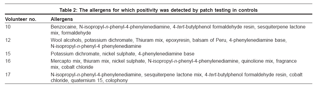
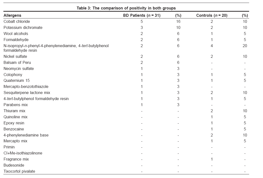
Indian Journal of Dermatology, Venereology and Leprology, Vol. 74, No. 2, March-April, 2008, pp. 187 Net letter Patch test in Behcet's disease Gül Ülker, Gönül Müzeyyen, Çakmak Seray Külcü, Kiliç Arzu Ankara Numune Education and Research Hospital, 2nd Dermatology Clinic, Ankara Code Number: dv08080 Sir, Behcet′s disease (BD) is a systemic inflammatory disease. Epidermal Langerhans cells (LCs) were shown to be activated in BD and the number of LCs is increased in BD. [1],[2] We investigated whether contact hypersensitivity is therefore increased in BD patients by patch testing. Thirty-one patients with BD who were diagnosed according to the criteria of the International Study Group for BD were enrolled in this study. Twenty age- and sex-matched individuals were selected to be a control group. European standard series (Chemotecnique Diagnostic TM ) was used for patch testing. The results were evaluated after 48, 72 and 96 hours. Marked erythema, edema and vesicle formation in any of the evaluations were accepted as a positive reaction. The ages of the patients with BD were between 18 and 56 years (mean 34.2 ± 1.6 years). There were 11 males and 20 females. Eleven (35.5%) patients tested positive to one or more allergens. In the control group, five individuals (25%) tested positive to one or more allergens [Table - 1],[Table - 2],[Table - 3]. When the positivity to allergens in patients with BD were compared with positivity in controls using the chi-square test, no significant difference was found ( P = 0.431) [Table - 3]. When the positivity for each allergen was compared within the patients′ and the control groups by the chi-square test, no statistically significant difference was found. Behcet′s disease (BD) is recognized as a systemic inflammatory disease of unknown etiopathogenesis. [3],[4] Langerhans cells (LCs) are situated suprabasally in most of the stratified squamous epithelia such as the epidermis and the epithelium of oral mucosa. They are thought to act as antigen-presenting cells during the induction of immune responses and because of this, they play an important role in contact hypersensitivity. There is an increase in the number of LCs in the skin of BD patients. [1],[2],[5] Kohn et al. showed that LCs were situated in the middle and upper parts of the epidermis and that LCs were bigger in BD patients, with prominent, well-developed, rough endoplasmic reticulum. These LCs in BD patients had significantly more granules and Kohn et al. suggested that this might be an expression of the active state of LCs and that LCs might be part of the complex pathogenesis of BD. [1] The findings of the studies of Kürkηüoπlu et al. and Lombardi et al. also supported this hypothesis. [2],[5] The exact role of LCs in BD is still unknown. In this study, we investigated whether contact hypersensitivity increases in BD. There are no similar studies in BD patients, as far as we know; this study is the first study in which patch testing was performed in BD patients. In our study, we found positivity mostly to cobalt chloride (five patients) and potassium dichromate (three patients). Positive reactions were detected in 35.5% in BD patients but only in 25% of the individuals in the control group. However, no significant difference was found when the positivity to allergens in BD patients and control individuals were compared. Also, no significant difference was found when the positivity for each allergen was compared within the patient and control groups. These findings suggest that contact hypersensitivity does not change in BD patients. We performed patch testing with European standard series. Significant outcomes might be observed with wider series in further studies. References
Copyright 2008 - Indian Journal of Dermatology, Venereology and Leprology The following images related to this document are available:Photo images[dv08080t1.jpg] [dv08080t3.jpg] [dv08080t2.jpg] |
| |||||||||
