 |
| About Bioline | All Journals | Testimonials | Membership | News |
|
||||||
|
||||||
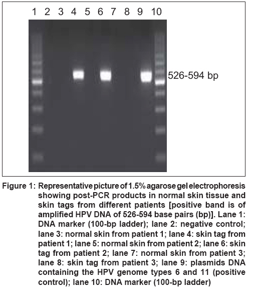
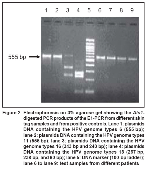
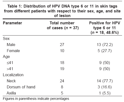
Indian Journal of Dermatology, Venereology and Leprology, Vol. 74, No. 3, May-June, 2008, pp. 222-225 Original Article Human papillomavirus and skin tags: Is there any association? Gupta Sachin, Aggarwal Ritu, Gupta Somesh, Arora SunilK Department of Immunopathology, Postgraduate Institute of Medical Education & Research, Chandigarh Code Number: dv08091 Abstract Background: Low-risk human papillomavirus (HPV) infections are related to the genesis of various benign lesions. In an isolated report available, HPVs have been implicated in the causation of skin tags too.Aims: The present study was designed to detect the existence of low-risk HPV types 6 and 11 in cutaneous soft fibromas (skin tag) in north Indians. Methods: A total of 37 cases of skin tags from various sites were analyzed. Highly sensitive and comprehensive polymerase chain reaction (PCR) and restriction fragment length polymorphism (RFLP) assays were done for the detection of low-risk HPV types 6 and 11. Results: The results revealed the presence of HPV DNA 6/11 in 48.6% of the skin tags examined by PCR-RFLP. Conclusion: This result corroborates the hypothesis that HPV plays a part in the etiology of benign lesions like cutaneous soft fibromas. The identification of HPV 6/11 in these lesions, which are benign proliferations of the skin, further expands the spectrum of HPV-linked lesions. Keywords: Human papilloma virus, Skin tags Introduction Human papillomavirus (HPV) is known to be the most ubiquitous of the human viruses. Over 100 HPV types have been identified till date. In healthy population, most of these HPV types appear to establish a latent infection of the skin, mostly as normal flora residing in hair follicles. The numerous HPV types differ in their biological properties and oncogenic potential. Types with high oncogenic potential (which always express the early proteins E6 and E7) are able to transform keratinocytes on their own. [1] The HPV genome usually remains episomal; but in transformed cells, the viral DNA is frequently integrated into the host DNA. [1] Skin tag, or soft fibroma, is a common benign condition, which consists of a bit of skin which projects from the surrounding skin. [2] Histologically, skin tag is a polypoid lesion with overlying mildly acanthotic epidermis. There is a loose, edematous fibrovascular core with mild chronic inflammation. Fibroepithelial polyps, or acrochordons, often develop in areas of skin friction. Certain HPV types are found to be associated with the pathogenesis of benign lesions like papillomas of larynx, conjunctiva; respiratory papillomatosis [3] ; etc. We undertook the present study with the aim of establishing the presence of low-risk human papillomavirus types 6 and 11 using polymerase chain reaction (PCR) systems in a benign cutaneous lesion like skin tag in a tertiary care hospital setting in India. Methods Those subjects who presented to the dermatology outpatient clinic with skin tags on multiple sites and were otherwise healthy and willing to participate in the study were enrolled. After obtaining informed consent, biopsy specimens from skin tags were obtained from neck, dorsum of hand and axilla from 37 patients. Ten biopsy specimens from the normal skin surrounding skin tags were also analyzed. In a few cases, biopsies from two different skin tags of the same patient were analyzed. DNA extraction E1-PCR analysis Ten minutes of denaturation at 94°C for the first cycle, followed by 1 min each of denaturation at 94°C, annealing at 48°C, and extension at 72°C for 33 cycles was done. The last cycle was extended for 10 min at 72°C. The electrophoresis of amplified products was done, and the gel was stained with 0.5 µg/mL ethidium bromide to visualize the amplified PCR product. A 526-594-base pair (bp) band was visualized in the samples positive for HPV on a UV transilluminator. The picture was captured on a gel documentation system (Imagemaster, Pharmacia Biotech, Sweden). Restriction fragment length polymorphism (RFLP) Statistical analysis Results Thirty-seven cases of skin tags from different sites including neck, dorsum of hand, and axilla were recruited in the study. Male:female ratio was approximately 3:1. Majority of the patients (57%) were in the age group of 26 to 55 years. Mean age was 41 years (range, 15-65). PCR using consensus primers spanning the E1 open reading frame showed presence of mucosotropic HPV types in 48.6% (18/37) of the samples [Figure - 1]. The biopsy specimens from the normal skin surrounding skin tags were negative for HPV DNA [Figure - 1]. The E1-PCR products were then subjected to RFLP, which specifically identifies HPV types 6 and 11. All samples (18/18, 100%) positive by E1-PCR showed HPV 6/11 sequence patterns (a single band of 555 bp) ([Figure - 2], lane 6 to lane 9). A few cases, where biopsies from two different sites of the same patient were analyzed, gave the same results. [Table - 1] shows the distribution of HPV DNA types 6 or 11 in skin tags from different patients with respect to their sex, age, and localization of the lesion. There was no significant correlation with respect to sex, site of lesion, or age of the patient. Discussion The subtropical and tropical areas of the globe harbor large number of infections. The reason can be attributed to the intense exposure of skin to sun. The ultraviolet (UV) irradiation has intense effects on skin immunology, including reduction in density and antigen-presenting ability of Langerhans cells in the epidermis, increased keratinocyte secretion of IL-10 and prostaglandin E2 with increased serum levels of IL-4. [5] In addition, there is induction of suppressive IL-12p40 homodimers by dendritic cells and macrophages. Thus UV irradiation seems to induce a resultant immunosuppressive effect with decreased TH1 cell activation. [6] This type of microenvironment is ideal for the survival of infection in healthy subjects living in tropical parts of the globe. HPVs are epitheliotropic and host specific, with infection across the species being exceedingly uncommon. It has been postulated that HPV infection begins with the inoculation of virus into the interrupted epithelium and the interaction with a putative specific cellular receptor. [7] It is recognized that HPV following trauma of epithelium establishes a nonproductive infection of basal cells in the skin and mucosa, but it is only in the differentiated epithelia that HPV replicates. The skin tags or fibroepithelial polyps are known to develop in areas of skin friction, leading to disruption of skin, which might serve as a route of entry for the virus. The presence of HPV DNA and mechanical friction seem to be significant cofactors in the pathogenesis of skin tags. [8] The immune status and genetic profile of the host, as well as the type of virus, may play a role in determining the clinical outcome of HPV infection. So the clinical behavior of soft fibromas may be reminiscent of that of recurrent laryngeal papillomas with respect to the fact that they spread locally in the same subject but rarely to other individuals. In the index study, HPV 6/11 DNA was found to be present in 48.6% of biopsies from skin tags. The entire samples were subjected to PCR-RFLP, and the results of both recognized the presence of HPV 6/11 in 48.6% of the entire sample. The samples were negative for high-risk HPV types (data not shown) when subjected to polymerase chain reaction using consensus primers for high-risk types. The only available literature is a study by Dianzani et al., [8] who have reported the presence of HPV 6/11 in 88% of the skin tags in Caucasian patients using PCR-RFLP technique. These observations strongly suggest that HPV, along with other cofactors, may be involved in the pathogenesis of these cutaneous lesions. Dianzani et al. [8] observed low quantity of HPV DNA in soft fibromas and explained their association with the clinical evolution of these lesions. The consistent presence of certain ′low-risk′ human papillomavirus types in the skin tag specimens from different sites of the same patient supports the viral etiology. Though the tropical geographical conditions favor infectious etiology in our patients, the lower frequency of the presence of HPV DNA in soft fibroma cells in the present study as compared to the frequencies reported in the literature could be due to difference in the sensitivity of the test, as well as the loss of HPV genome. The presence of HPV sequences in skin tags could aid their recurrence as well. The expression of early viral genes may contribute to stimulated cell growth, which leads to limited epithelial proliferation and formation of acanthotic epidermis overlying edematous fibrovascular tissue. The association of HPV type 6 with benign lesions is very old. It was in 1982 that HPV type 6 was observed in laryngeal papillomas with the help of DNA hybridization technique. A year later, HPV type 11 was also found to be associated with laryngeal papillomas. [9] However, the literature on the intracellular mechanisms through which HPV type 6 or 11 may immortalize cells is still fractional in comparison with data on the high-risk HPV types. Additional in vitro and epidemiological studies investigating the influence of genetic and environmental factors on the interaction of the HPV proteins with cellular proteins should provide valuable information on a possible role for HPV types 6 and 11 in the pathogenesis of these cutaneous lesions. Acknowledgment We are grateful to Dr. Ethel-Michele de Villiers, Referenzzentrum fur humanpathogene papillomviren, Abteilung Tumorvirus-Charakterisierung, Heidelberg, Germany, for providing plasmid DNA of HPV types 6, 11, 16, and 18.References
Copyright 2008 - Indian Journal of Dermatology, Venereology and Leprology The following images related to this document are available:Photo images[dv08091f2.jpg] [dv08091f1.jpg] [dv08091t1.jpg] |
| |||||||||
