 |
| About Bioline | All Journals | Testimonials | Membership | News |
|
||||||
|
||||||
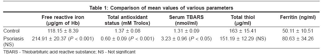
Indian Journal of Dermatology, Venereology and Leprology, Vol. 74, No. 3, May-June, 2008, pp. 277-278 Letter To Editor Role of free reactive iron in psoriasis Ghosh Arpita, Mukhopadhyay Soma, Kar Manoj Department of Biochemistry, NRS Medical College and Hospital, Kolkatta, West Bengal Code Number: dv08116 Sir, Psoriasis is a chronic, inflammatory autoimmune skin disease. [1] Recently, oxidative stress has been implicated in the etiopathology of psoriasis. Reduced level of serum super oxide dismutase (SOD), glutathione peroxidase (GPX), and elevated level of nitric oxide (NO) has been reported. [2],[3],[4] The causative factor of such oxidative stress is not yet clearly known. In psoriatic plaque blood capillaries are dilated and become tortuous to form loops which may cause breakdown of erythrocytes to release hemoglobin. Moreover, low GPX and SOD may help to elevate the level of hydrogen peroxide (H 2 O 2 ) which further causes break down of hemoglobin within erythrocyte to form nonheme reactive iron. This free reactive iron can catalyze Haber-Weiss reaction and generate deadly damaging hydroxyl radical which in turn damage cellular constituents. [5] To investigate whether free reactive iron has any role in secondary pathogenesis of psoriatic lesion, we sought to study the level of free reactive iron in stroma free hemolysate. Serum level of cellular damage marker thiobarbituric acid reacting substance (TBARS), total thiol, total antioxidant status (TAS), and ferritin were also measured from blood sample of psoriasis patients by standard methods [Table - 1]. Sixteen patients were selected for our study (13 males and 3 females) and all had stable plaque type psoriasis, two with psoriasic arthritis. Patients were within the age group of 25-36. The psoriasis area severity index of the patients ranged from 10 to 60. The mean level of free reactive iron from stroma free hemolysate (214.91 μgm/g of Hb) as compared to that of normal control volunteers (118.15 μg/g of Hb) and the cellular damage marker TBARS (3.23 nmol/ml) compare to that of normal control volunteers (1.31 nmol/ml) were found to highly significant ( P < 0.001) and significant ( P < 0.05), respectively. The TAS level was also found to be significantly low (0.6 Trolox equivalent) when compare to normal control (1.37 Trolox equivalent). The values of total thiol and ferritin were not statistically significantly different, but mean values were lower for total thiol and higher for ferritin. Ferrous iron with six coordination states is bound and tamed within protoporphyrin ring of hemoglobin. Under specific circumstances like oxidative stress or over production of H 2 O 2 , it comes out of the ring and can ligate with other part of globin chain. This non-heme iron has been termed free reactive iron which can generate cytotoxic hydroxyl radical by Fenton reaction in presence of H 2 O 2 . [5] Our findings indicate the increase in level of free reactive iron and lower level of antioxidant status in psoriasis. This may contribute via free radical generation to the development of secondary cellular damage and pathological state in psoriasis vulgaris. Therapeutic use of iron chelator and antioxidant drugs may be investigated for beneficial role in psoriasis. References
Copyright 2008 - Indian Journal of Dermatology, Venereology and Leprology The following images related to this document are available:Photo images[dv08116t1.jpg] |
| |||||||||
