
|
Indian Journal of Dermatology, Venereology and Leprology
Medknow Publications on behalf of The Indian Association of Dermatologists, Venereologists and Leprologists (IADVL)
ISSN: 0378-6323 EISSN: 0973-3922
Vol. 74, Num. 4, 2008, pp. 311-321
|
Indian Journal of Dermatology, Venereology and Leprology, Vol. 74, No. 4, July-August, 2008, pp. 311-321
Review Article
Adult onset pityriasis rubra pilaris
Sehgal VirendraN, Srivastava Govind, Dogra Sunil
Dermato-Venereology (Skin/VD) Centre, Sehgal Nursing Home, Delhi
Correspondence Address:A/6, Panchwati, Delhi-110 033
drsehgal@ndf.vsnl.net.in
Code Number: dv08148
Abstract Pityriasis rubra pilaris (PRP) has always been an intriguing topic ever since its inception. It is a group of chronic disorders characterized by reddish orange plaques with pityriasiform scaling showing follicular keratoses, palmoplantar keratoderma, and sometimes, erythroderma. It occurs all over the world but with racial variations. Its incidence might vary and the age at onset, behavior, clinical appearance, and prognosis are considered to be very important for its classification. It may manifest either as Type I classical adult onset PRP, Type II atypical adult (onset) PRP, or Type VI PRP (HIV-associated PRP pityriasis rubra pilaris) in contrast to classical juvenile (Type III) and circumscribed juvenile (Type IV) encountered among children. Its diagnosis is largely clinical with microscopic pathology being a useful supplement, but it continues to be a therapeutic dilemma. We review the epidemiology of adult onset PRP here and take stock of the prevalent treatment options.
Keywords: Adult onset, Pityriasis rubra pilaris
Introduction Ever since the first reported case of the disease, pityriasis rubra pilaris (PRP) has remained a consistently recorded and researched entity to date. [1],[2],[3],[4],[5],[6],[7],[8],[9],[10],[11],[12] However, its etiology and management have remained a challenge for the treating physician. It is seen in adults (adult onset) as well as in children, and affects both the sexes. Occasionally, PRP is associated with other diseases and it was speculated that the disorder might be the result of an abnormal immune response to some antigenic stimuli. [4] However, familial occurrence of the disease might point to some genes that predispose the individual to develop this disorder after certain precipitating events. [13],[14],[15],[16],[17] The occurrence of this dermatosis in association with human immunodeficiency virus (HIV)/acquired immunodeficiency disease (AIDS) patients has sparked a dialogue as to whether or not it is yet another variant of PRP. [18],[19],[20],[21],[22]
Definition Pityriasis rubra pilaris refers to a group of chronic disorders characterized by reddish orange plaques with pityriasiform scaling showing follicular keratoses, palmoplantar keratoderma, and sometimes, erythroderma. Familial as well as acquired forms of the disease have been reported. [23]
History Devergie has been credited with the naming of ′pityriasis pilaris′ in 1857, which received the eponym of Devergie′s diseases. [24] However, much before that, Tarral [1] in 1835, recorded the case description of this disease in the "Rayer′s [1] -a theoretical and practical treatise on the disease of the skin" under the title of ′general psoriasis′. Devergie stressed that Tarral′s case was an example of PRP, and observed that PRP might be confused with psoriasis. However, in 1889, Besnier [25] advanced the present day name, "pityriasis rubra pilaris." Later, several authors [26],[27],[28],[29],[30],[31] recognized that PRP was of many types, and thus suggested several working classifications. [32],[33],[34],[35],[36],[37] With the advent of HIV/AIDS, its association with PRP has been noticed by many, prompting expansion of the existing classification to accommodate PRP associated with HIV as a distinct type. [20],[38],[39],[40]
Epidemiology Although PRP occurs worldwide, there are racial variations. [2],[3] Its incidence might vary-it is 1 in 5,000 in Great Britain [34] and 1 in 50,000 in India [31] in an outpatient setting. Both the sexes are affected equally at all ages. [14],[33] A bimodal or trimodal age distribution has been recorded with peak incidence in the 1 st , 2 nd and 6 th decade of life. [27],[34],[41],[42],[43],[44] The majority of the cases have been acquired [34],[35],[36],[37],[38],[39],[40],[41],[42] and familial occurrence is only sporadic (up to 6.5%). [13],[14],[15],[16],[17],[27],[28],[37],[41],[45],[46] Autosomal dominant inheritance with variable penetrance is usual; however, autosomal recessive inheritance has also been described. [47] Monozygotic twins have been observed to develop PRP. [48] Familial PRP usually develops in childhood while acquired PRP develops in the 5 th or 6 th decade of life. [32],[33],[34] The development of PRP in HIV/AIDS was recognized several years after its discovery, and might show peculiarities compared to classical adult PRP; it responds to antiretroviral therapy in most instances.
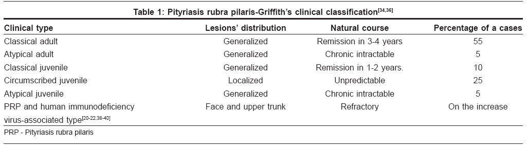
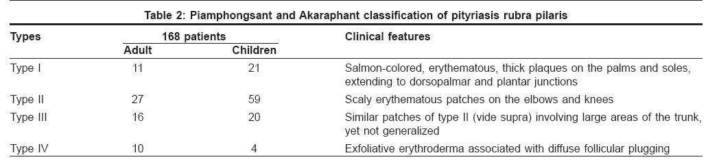
Classification PRP was initially classified on the basis of the age at onset, behavior, clinical appearance, and prognosis by Griffiths [34] in 1980 [Table - 1]. The classical (type I) adult onset PRP shows a characteristic morphology and usually resolves in 3-4 years, whereas atypical adult-onset (type II) PRP is chronic, shows ichthyosiform and lamellar scales on the palms and soles, and alopecia of varying degrees. [2],[34] The association of PRP and HIV infection has recently been identified as type VI PRP and most of the cases have been reported in young heterosexual/homosexual men. [22] It has characteristically nodulo-cystic and lichen spinulosus-like lesions, poor prognosis, and is refractory to treatment. [13],[14],[15],[18],[19],[20],[22],[28] However, after the study of 168 Thai patients, Piamphongsant and Akaraphant [37] classified the disease into four types based on the physical findings [Table - 2]. However, Griffiths′ [34] classification continues to be the mainstay in practice for delineating the disease.
Etiology
The exact cause of PRP is not known-the familial type usually has an autosomal dominant mode of inheritance, [46] although recessive forms have also been recorded. [47] Genetic factors may be important; however, family history is generally not forthcoming. Epidermal hyperactivity demonstrated by a faster growth of the nails and an increase in the thymidine labeling index from a normal 3% to a high 27%, may be observed in PRP. [49],[50],[51],[52],[53] Finzi et al, observed a decreased level of serum retinol-binding protein in 11 PRP patients and their relatives, [54],[55],[56],[57],[58] while Frazier and Hu [59],[60] and Lowenthol [61] suggested that an abnormal vitamin A metabolism and/or vitamin A deficiency may play some role in PRP etiology. However, others [62],[63],[64] did not find any decreased levels of vitamin A; thus, no correlation between vitamin A deficiency and dyskeratosis has been established. [65] Interestingly, Rothman observed that vitamin A administration has often been beneficial in follicular and nonfollicular, hyperkeratotic disease even if these diseases did not originate from vitamin A deficiency. [66]
Furthermore, bacterial superantigens have recently been incriminated in triggering some skin diseases including juvenile PRP. [67],[68],[69],[70] This has been corroborated by the detection of bacterial superantigens in the course of acute throat infections ( Staphylococcus aureus and group A b S treptococcal pyogenes ), simultaneous appearance of lesions conforming to the morphology of childhood onset/juvenile PRP, and the disappearance of lesions following administration of appropriate antibiotics. In addition, significant increases in peripheral blood mononuclear cell (PBMCs) counts against Staphylococcal enterotoxin B in vitro might suggest hyper-reactivity to some bacterial products, which may lead to childhood onset/juvenile PRP. [68]
Clinical Features
Adult onset PRP conforms to Griffiths [34] type I classical adult and type II atypical adult classifications, the former being the most common. In contrast to childhood onset juvenile PRP, [23] adult PRP typically starts on the face and scalp and promptly spreads in the cephalocaudal direction. [2],[3]
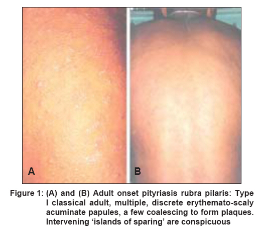
Type I classical adult onset PRP: It is characterized by follicular hyperkeratotic papules that coalesce into large, scaly, erythematous plaques, palmoplantar keratoderma, diffuse furfuraceous scaling of the scalp sometimes progressing into erythroderma. [2],[3],[4],[24],[25],[26],[27],[28] The onset is usually acute and the eruptions begin on the head, neck and upper chest as discrete, follicular papules that often coalesce to form plaques with interfollicular erythema. The spread of the lesion is characteristically in the cephalocaudal direction. The face assumes a red-orange hue with mild to moderate ectropion. The affected skin is extremely rough to touch and feels like a file. [1] Prolonged erythema may cause resultant edema, and may precipitate a high output cardiac failure in the elderly. The palms and soles may acquire the appearance of a ′hyperkeratotic sandal′, while the scalp reveals diffuse bran-like scaling. Should an erythroderma develop, a few sharply demarcated islands of unaffected skin [Figure 1A, B] are important diagnostic criteria. [2],[3],[71],[72],[73],[74],[75],[76] Pruritus is uncommon; nail changes (if any) are marked by thickening and yellow-brown discoloration of the nail plate, subungual hyperkeratosis, and splinter hemorrhages. [2],[3],[77],[78],[79] Unlike psoriasis, nail dystrophy and pitting are minimal in PRP. The oral mucosa may be involved in a few patients, showing macular erythema, diffuse hyperkeratosis, and white streaks; [80] hair and teeth are normal. [2],[3],[4] Type I classical adult onset PRP runs a chronic course, three out of four cases may resolve in 1-3 years; relapses are usually uncommon.
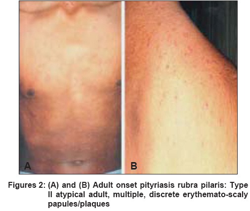
Type II atypical adult onset PRP: It is an uncommon form of the disease that develops in middle-aged adults with atypical morphological features deviating from those described above . These patients show an admixture of follicular hyperkeratosis and lamellar scaling [Figure 2A, B] on their skin surface. [2],[3],[4],[24],[81] Areas of eczematous changes can sometimes confuse the clinical picture. The classical cephalocaudal progression is conspicuous by its absence; the occurrence of erythroderma is also unusual.
Type VI PRP (HIV-associated PRP): The occurrence of PRP in HIV/AIDS shows certain peculiarities [20],[38],[39],[40] such as a ′filiform′ pattern of keratosis on the face and upper trunk, accompanied by marked acne conglobata. This type is usually recalcitrant to conventional therapy and has a poor prognosis. [20],[21],[22],[38],[39],[40] Other types are described in [Table - 1].
Associated Findings Adult PRP has been found to be associated with several cutaneous and noncutaneous disorders. [82],[83],[84],[85] the exact significance of which is a matter of speculation. The associated disorders include vitiligo, lichen planus, alopecia universalis, [12] Kaposis varicelliform eruption, [86] sero-negative arthritis, [87],[88],[89],[90] myositis, [83] myasthenia gravis, [91] hypothyroidisim, [82] celiac sprue, [84] and other infections including HIV. [20],[38],[39],[40] Infrequently, internal malignancies have been recorded in adult onset PRP. [92],[93],[94],[95] However, prominent or increasing seborrhoeic keratoses seen in erythrodermic PRP does not necessarily imply an underlying malignancy. [85],[96] An intense degree of erythema in erythrodermic PRP predisposes the individual to photosensitivity and worsening of the erythema has also been recorded with UVA and UVB. [97],[98],[99]
Histopathologic Findings
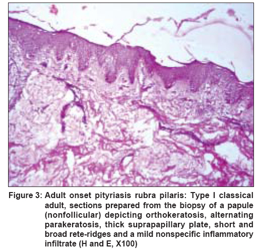
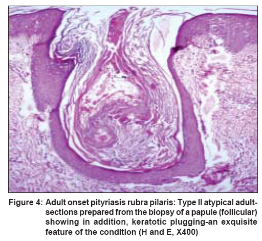
Adult onset PRP displays distinctive histopathological findings, which may differ according to the stage and evolution of the lesions. [2],[3] The salient criteria include: (a) alternating orthokeratosis and parakeratosis in both the vertical and horizontal directions, (b) hypergranulosis, (c), irregular acanthosis apparent in the form of short and broad rete-ridges, (d) thick suprapapillary plates, and (e) a sparse to moderate lymphocytic perivascular infiltrate in the dermis [27],[42],[49],[71],[100] [Figure - 3]. The hair follicles are dilated and filled with a dense, horny plug [Figure - 4]. Munro′s microabscesses and suprapapillary thinning are conspicuously absent. The differential diagnosis may often be difficult in erythrodermic patients.
Walsh et al, [101] found that dermatopathologist are the least (25%) accurate when scanning the sections prepared from biopsies of erythroderma of PRP origin. The dermis shows dilated capillaries with a mild to moderate infiltrate of lymphocytes and histiocytes. Acantholysis and focal acantholytic dyskeratosis have recently been recorded in adult PRP. [94],[102],[103],[104] These histological parameters are unique and different from those seen in psoriasis; Magro and Crowson [43] found these features in 23 of the 32 biopsies from PRP. However, they were not found in any of the specimens of psoriasis.
Porter and Shuster [105] have been credited with the demonstration of increased epidermal replacement after they found an increase in the uptake of amino acids by the PRP lesions. Later on, several in vivo and in vitro autoradiography studies using titrated thymidine confirmed an increase in the labeling index of PRP epidermal cells when compared with normal, reflecting increased cell proliferation. [42],[49],[50],[51],[53],[106] Electron microscopy revealed a decrease in the number of tonofilaments, desmosomes, and enlarged intercellular spaces. [49],[107] The corneocytes are fusiform and show numerous pits. [107] Evidence of parakeratosis of the stratum corneum is seen as lipid-like vacuoles, incomplete keratinization, and remnants of nuclei. [58],[107]
Laboratory Findings Hematological and laboratory test results are usually within normal limits; the main emphasis remains on the histopathology. Plasma vitamin A and carotenoid levels are normal, [62],[63],[64] although retinal-binding protein may be low [54] or normal. [55],[56],[57],[58] Direct immunofluorescence tests with antibodies to human IgG, IgM, IgA and complement C 3 were found to be negative in 15 adult PRP patients by Niemi et al. [42] However, immunoelectrophoresis of the scales from PRP demonstrated the presence of only IgG, while psoriatic scales had IgG, IgA and C 3.[108] Takematsu et al, [109],[110] recorded normal levels of leukotriene B4 but low levels of anaphylotoxins in scale extracts from PRP. Other studies have been done on HLA typing, [42] direct immunofluorescence, [42] keratin monoclonal antibodies, [15] parathyroid hormone levels, [111] and a western blot analysis of the skin, [15] but these studies only have academic significance.
Diagnosis Until the disease is well-developed, it may be difficult to diagnose with full confidence. However, repeated observations and a few biopsies may confirm the diagnosis. [2],[3],[4],[24],[112],[113],[114],[115],[116] Atypical (type II) PRP may be more difficult to diagnose than classical adult onset (type I) PRP. Follicular hyperkeratosis on the back of the fingers, orange-colored eruptions with intervening areas of normal skin ′islands of sparing′ and/or palmoplantar keratoderma are features of classical adult onset PRP; they are ill defined in atypical adult PRP. [2],[3],[4],[117] The differential diagnosis of adult onset PRP usually includes psoriasis. [2],[4],[118] The absence of Auspitz and candle grease signs is an instant clinical diagnostic clue. [2],[3] Erythrodermic PRP can be confused with other forms of erythroderma [119] and skin biopsy of such patients can confirm PRP only on an exclusion basis. [101] Arthropathic PRP is unusual to record. [120] Resolving PRP may mimic seborrhoeic dermatitis [2] or erythema gyratum repens. [17],[121] PRP may be a cutaneous marker of internal malignancy, [92],[93],[95],[122] leukemia, [93] metastatic carcinoma, [92] or Sezary syndrome [123] in adults, which may follow after a variable length of time. Interestingly, cutaneous T-cell lymphoma and Sezary′s syndrome also form a differential diagnosis of erythrodermic PRP. [124],[125] Rarely, dermatomyositis may develop skin eruptions akin to the adult onset PRP. [126],[127],[128],[129],[130] Heteroduplex analysis of T-cell receptor gamma gene arrangements may be a newer adjuvant diagnostic tool in skin biopsies from erythrodermas. [128]
Treatment Options
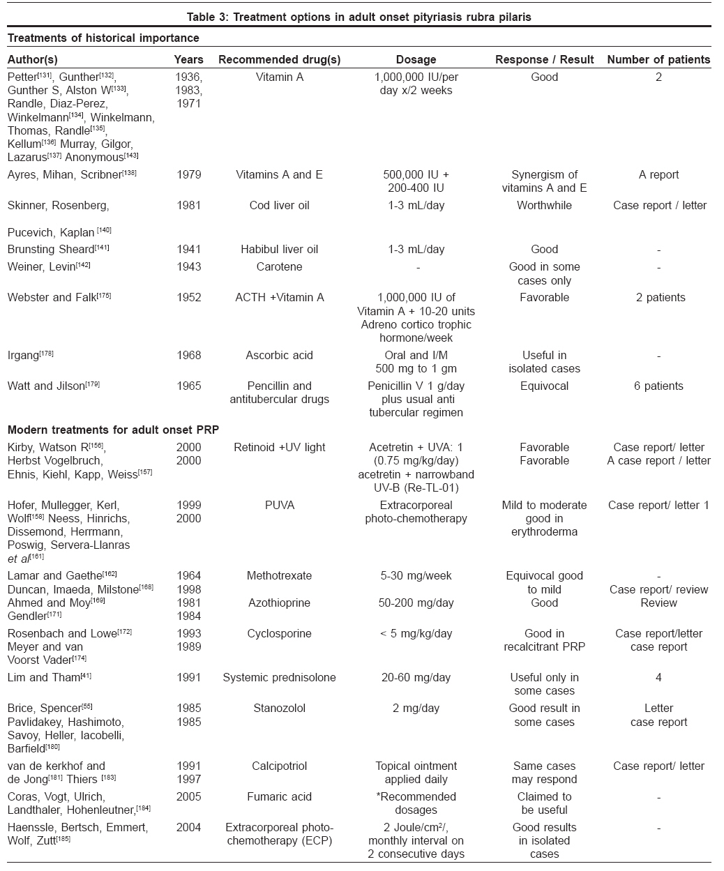
The diagnosis and treatment of PRP have always been a source of great interest. There is no acclaimed treatment for PRP at present. Thus, affected individuals often visit and change many treating dermatologists to alleviate their signs and symptoms. More often than not, it is an exercise in futility as the treating physician/dermatologist too is in dilemma. Several treatment [131],[132],[133],[134],[135],[136],[137],[138],[139],[140],[141],[142],[143],[144],[145],[146],[147],[148],[149],[150],[151],[152],[153],[154],[155],[156],[157],[158],[159],[160],[161],[162],[163],[164],[165],[166],[167],[168],[169],[170],[171],[172],[173],[174],[175],[176],[177],[178],[179],[180],[181],[182],[183],[184],[185],[186],[187],[188],[189],[190],[191] options have been in vogue and are tabulated below [Table - 3].
Narrowband UV-B with oral retinoids has been useful in some cases. [182],[187] Topical calcipotriol [138] and tacalcitol have also given promising results in some patients. HIV-associated PRP is more recalcitrant but antiretroviral drug therapy has caused alleviation of the symptoms and may even cause complete regression in such patients. [188] Methotrexate has been found to be moderately effective. In an attempt to explore an ideal therapy, newer treatment options like biologicals (infliximab), calineurin inhibitors (pimecrolimus) etc. are being tried in PRP. [192],[193] The use of emollients to symptomatically improve the condition may also be useful. It is imperative to record at this point in time, that several treatment options that have been used so far may not be satisfactory as no organized drug trials are available. Nevertheless, isotretinoin, a retinoid, seems to be a plausible option. [149],[154],[189],[190],[191]
The historical and epidemiological perspectives of adult onset PRP as well as its etiology have been described. Microscopic pathology and its variations have been clearly defined, emphasizing its role in supplementing clinical diagnosis and treatment has been facilitated by the inclusion of a table for decision-making.
References
| 1. | Tarral C. General psoriasis-desquamation from the parts covered by hair. In: Rayer P, editor. A theoretical and practical treatise on the diseases of the skin. 2 nd ed, London: Baillere; 1835. p. 648-9. Back to cited text no. 1 |
| 2. | Griffiths WA, Judge MR, Leigh IM. Disorders of keratinization-pityriasis rubra Pilaris. In: Champion RH, Burton JL, Burns DA, et al, editors. Text Book of dermatology, 6 th ed. London: Blackwell Science; 1988. p. 1539-45. Back to cited text no. 2 |
| 3. | Gold Smith LA, Baden HP. Pityriasis rubra pilaris. In: Freeberg IM, Eisen AZ, Wolff K, et al, editors. Dermatology in general medicine. Vol 1 6 th ed. London: Mc-Graw Hill Publication; 2003. p. 442-4. Back to cited text no. 3 |
| 4. | Albert MR, Mackool BT. Pityriasis rubra pilaris. Int J Dermatol 1999;38:1-11. Back to cited text no. 4 |
| 5. | White KL. Pityriasis rubra pilaris. Dermatol Online J 2003;9:6. Back to cited text no. 5 |
| 6. | Vijayalakshmi AM, Malika A. Pityriasis rubra pilaris. Indian Pediatr 2003;40:432-3. Back to cited text no. 6 |
| 7. | Selvaag E, Haederstel M, Thomsen K. Pityriasis rubra pilaris: A retrospective study of 12 patients. J Eur Acad Dermatol Veneraol 2000;14:514-5. Back to cited text no. 7 |
| 8. | Sehgal VN, Bajaj P, Jain S. Pityriasis rubra pilaris -report of four cases. J Dermatol 2000;27:174-7. Back to cited text no. 8 |
| 9. | Sorensen KB, Thestrup-Pedersen K. Pityriasis rubra pilaris: A retrospective analysis of 43 patients. Acta Derm Venereol 1999;79:405-6. Back to cited text no. 9 |
| 10. | Varma S. Logan RA. Exanthematic pityriasis rubra pilaris. Br J Dermatol 1999;141:769-71. Back to cited text no. 10 |
| 11. | Clayton BD, Jorizzo JL, Hitchcock MG, Fleischer AB Jr, Williford PM, Feldman SR, et al. Adult pityriasis rubra pilaris: A 10 year case series. J Am Acad Dermatol 1997;36:959-64. Back to cited text no. 11 |
| 12. | Cecchi R, Giomi A, Tuci F, Bartoli L, Seghieri G . Pityriasis rubra pilaris, lichen planus, alopecia universals and vitiligo in a single patients with chronic viral hepatitis C. Dermatology 1994;188:239-40. Back to cited text no. 12 |
| 13. | Griffiths A. Pityriasis rubra pilaris - Etiologic considerations . J Am Acad Dermatol 1984;10:1086-8. Back to cited text no. 13 |
| 14. | Sehgal VN, Jain S, Kumar S, Bhattacharya SN, Sardana K, Bajaj P. Familial pityriasis rubra pilaris (Adult classic - I): A reports of 3 cases in a single family. Skinmed Dermatol clin 2002;1 : 161-4. Back to cited text no. 14 |
| 15. | Vanderhooft SL, Francis JS, Holbrook KA, Dale BA, Fleckman P . Familial pityriasis rubra pilaris. Arch Dermatol 1995;131 : 448-53. Back to cited text no. 15 |
| 16. | Holden CA, Curley RK. Down's syndrome and pityriasis rubra pilaris. Clin Exp Dermatol 1989;14 : 332. Back to cited text no. 16 |
| 17. | Hazini AR, Rongioletti F, Rebora A. Pityriasis rubra pilaris and vitiligo in Down's syndrome. Clin Exp Dermatol 1988;13 : 334-5. Back to cited text no. 17 |
| 18. | Bonomo RA, Korman N, Nagashima-Whalen L Briggs J, Graham R, Salata RA . Pityriasis rubra pilaris: An unusual cutaneous complication of AIDS. Am J Med Sci 1997;314 : 118-21. Back to cited text no. 18 |
| 19. | Misery I, Faure M, Claidy A. Pityriasis rubra pilaris and HIV infection- type 6 pityriasis rubra pilaris. Br J Dermatol 1996;135:1008-9. Back to cited text no. 19 |
| 20. | Miralles ES, Nunez M, De Las Heras ME, Perez B, Moreno R, Ledo A . Pityriasis rubra pilaris and HIV infection. Br J Dermatol 1995;133 : 990-3. Back to cited text no. 20 |
| 21. | Resnick SD, Murrel DF, Woosley JT. Pityriasis rubra pilaris, acne conglobata, and elongated follicular spines: An HIV associated follicular syndrome. J Am Acad Dermatol 1993;29 : 283. Back to cited text no. 21 |
| 22. | Blauvelt A, Nahass GT, Pardo RJ, Kerdel FA . Pityriasis rubra pilaris and HIV infection. J Am Acad Dermatol 1991;24 : 703-5. Back to cited text no. 22 |
| 23. | Sehgal VN, Srivastava G. (Juvenile) pityriasis rubra pilaris. Int J Dermatol 2006;45:438-46. Back to cited text no. 23 |
| 24. | Devergie A. Traitι pratique des maladies de la peau. (In French) Paris: Masson; 1857. p. 454-64. Back to cited text no. 24 |
| 25. | Besnier E. Observation pour server a historic clinique du, pityriasis rubra pilaris (In French). Am Dermatol Syphil ( Paris ) 1889;10:253-87. Back to cited text no. 25 |
| 26. | Bergeron JR, Stone OJ. Follicular occlusion triad in a follicular blocking disease (pityriasis rubra pilaris). Dermatologica 1968;136 : 362-7. Back to cited text no. 26 |
| 27. | Davidson CL Jr, Winkelman RK, Kierland RR. Pityriasis rubra pilaris: A follow up study of 57 patients. Arch Dermatol 1969;100 : 175-8. Back to cited text no. 27 |
| 28. | Kint A, De Bie S, Geerts ML, T'Kint R . Pityriasis rubra pilaris: A familial condition. Arch Belg Dermatol Syphilgr 1972;28 : 371-6. Back to cited text no. 28 |
| 29. | Binnick SA. Pityriasis rubra pilaris. Int J Dermatol 1979;18 : 587-8. Back to cited text no. 29 |
| 30. | Fox BJ, Odom RB. Papulo - squamous disease: A review. J Am Acad Dermatol 1985;12 : 597-624. Back to cited text no. 30 |
| 31. | Sehgal VN, Jain MK, Mathur RP. Pityriasis rubra pilaris in Indians. Br J Dermatol 1989;121 : 821-2. Back to cited text no. 31 |
| 32. | Griffiths WA. Pityriasis rubra pilaris: A historical approach. Trans St Johns Hosp Dermatol Soc 1975;61 : 58-69. Back to cited text no. 32 |
| 33. | Griffiths WA. Pityriasis rubra pilaris: An historical approach 2, Clinical features. Clin Exp Dermatol 1976;1 : 37-50. Back to cited text no. 33 |
| 34. | Griffiths WA. Pityriasis rubra Pilaris. Clin Exp Dermatol 1980;5 : 105-12. Back to cited text no. 34 |
| 35. | Gelmetti C, Cerri D. Pityriasis rubra pilaris-the problems of its classification. J Am Acad Dermatol 1990;23 : 1186-8. Back to cited text no. 35 |
| 36. | Griffiths WA. Pityriasis rubra pilaris-the problems of its classification. J Am Acad Dermtol 1992;26 : 140-2. Back to cited text no. 36 |
| 37. | Piamphongsant T, Akaraphant R. Pityriasis rubra pilaris: A new proposed classification. Clin Exp Dermtol 1994;19 : 134-8. Back to cited text no. 37 |
| 38. | Martin AG, Weaver CC, Cockerell CJ, Bergcr TG . Pityriasis rubra pilaris in the setting of HIV infection: Clinical behavior and association with explosive cystic acne. Br J Dermatol 1992;126 : 617-20. Back to cited text no. 38 |
| 39. | Sanchez-Regana M, Fuentes CG, Creus L, Salleras M, Umbert P . Pityriasis rubra pilaris and HIV infection: A part of the spectrum of HIV-associated follicular syndrome. Br J Dermatol 1996;135 : 1008-9. Back to cited text no. 39 |
| 40. | Gonzalez-Lopez A, Velasco E, Pozo T, Del Villar A . HIV- associated pityriasis rubra pilaris responsive to triple antiretroviral therapy. Br J Dermatol 1999;140 : 931-4. Back to cited text no. 40 |
| 41. | Lim JT, Tham SN. Pityriasis rubra pilaris in Singapore. Clin Exp Dermatol 1991;16 : 181-4. Back to cited text no. 41 |
| 42. | Niemi KM, Kousa M, Storgards K, Karvonen J. Pityriasis rubra pilaris: A clinico- pathological study with a special reference to autoradiography and histocompatibility antigens. Dermatologica 1976;152 : 109-18. Back to cited text no. 42 |
| 43. | Magro CM, Crowson AN. The clinical and histomorphological features of pityriasis rubra pilaris: A comparative analysis with psoriasis. J Cutan Pathol 1997;24:416-24. Back to cited text no. 43 |
| 44. | Jacky WK. Pityriasis rubra Pilaris in Black South Africans. Clin Exp Dermatol 1999;24 : 160-3. Back to cited text no. 44 |
| 45. | Gelmeti C, Schiuma AA, Cerri D, Gianotti F . Pityriasis rubra pilaris in childhood: A long term study of 29 cases. Pediatr Dermatol 1986;3 : 446-51. Back to cited text no. 45 |
| 46. | Betjkmann DB, Bith, Heuyer. Pityriasis rubra pilaris familial. Am Dermatol Syphiligr ( Paris ) 1910;1 : 609-19. Back to cited text no. 46 |
| 47. | Wells RS. In Discussion on Borrie P. Pityriasis rubra pilaris treated with Methotrexate. Br J Dermatol 1967;79 : 115-6 Back to cited text no. 47 |
| 48. | Knudsen EA. Pityriasis rubra pilaris in identical twins. Br J Dermatol 1958;70 : 27-9 Back to cited text no. 48 |
| 49. | Braun-Falco O, Ryckmanns F, Schmoeckel C, Landthaler M. Pityriasis rubra pilaris: A clinico pathological and therapeutic study with special reference to histochemistry, radiography and electron microscopy. Arch Dermatol Res 1983;275 : 287-95. Back to cited text no. 49 |
| 50. | Harper RA, Risplfr J. Pityriasis rubra pilaris epidermal cell in vitro : A comparison with normal and psoriatic cells. Arch Dermatol Res 1977;260 : 253-5. Back to cited text no. 50 |
| 51. | Ralfs IG, Dawber RP, Ryan TJ, Wright NA . Pityriasis rubra pilaris-epidermal cell kinetics Br J Dermatol 1981;104:249-52. Back to cited text no. 51 |
| 52. | Kanitakis J, Hoyo E, Chouvet B, Thivolet J, Faure M, Claudy A. Keratinocyte proliferation in epidermal keratinocyte disorders evaluated through PCNA/ Cyclin immunolabelling and AGNOR counting. Acta Derm Venerol 1993;73 : 370-5. Back to cited text no. 52 |
| 53. | Griffiths WA, Pieris S. Pityriasis rubra pilaris: An autoradiographic study. Br J Dermatol 1982;107 : 665-7. Back to cited text no. 53 |
| 54. | Finzi AF, Altomare G, Bergamaschini L, Tucci A. Pityriasis rubra pilaris and retinol binding protein. Br J Dermatol 1981;104 : 253-6. Back to cited text no. 54 |
| 55. | Brice SL, Spencer SK. Stanozolol in the treatment of pityriasis rubra pilaris. Arch Dermatol 1985;121 : 1105-6. Back to cited text no. 55 |
| 56. | Van Voorst Vader PC, Van Oostveen F, Houthoff HJ, Marrink J. Pityriasis rubra pilaris, Vitamin A and Retinol binding protein: A case study. Acta Derm Venereol 1984;64 : 430-2. Back to cited text no. 56 |
| 57. | Stoll DM, King LE Jr, Chytil F. Serum levels of retinol binding protein in patients with pityriasis rubra pilaris. Br J Dermatol 1983;108:375. Back to cited text no. 57 |
| 58. | Kanerva L, Lauharanta J, Niemi KM, th Lassus A . Ultrastructure of pityriasis rubra pilaris with observation during retinoid treatment. Br J Dermatol 1983;108 : 653-63. Back to cited text no. 58 |
| 59. | Frazier CN, Hu CK. Cutaneous lesions associated with a deficiency of vitamin A in man. Arch Intern Med. 1931;48 : 507-14. Back to cited text no. 59 |
| 60. | Frazier CN., Hu CK. Nature and distribution according to age of cutaneous manifestation of vitamin A deficiency: A study of 207 cases. Arch Dermatol Syphilol 1936;33 : 825-52. Back to cited text no. 60 |
| 61. | Lowenthal LJ. A new cutaneous manifestation in the syndrome of vitamin A deficiency. Arch Dermatol Syphilol 1933;28 : 700-8. Back to cited text no. 61 |
| 62. | Cornbleet T. Liver Vitamin A in Darier's and Davergies disease. J Invest Dermatol 1954;23 : 71-3. Back to cited text no. 62 |
| 63. | Gross DA, Landay JW, Newcomer VD. Pityriasis rubra pilaris-report of a case and analysis of the literature. Arch Dermatol 1969;99 : 710-6. Back to cited text no. 63 |
| 64. | Griffiths WA. Vitamin A and pityriasis rubra pilaris. J Am Acad Dermatol 1982;7 : 555. Back to cited text no. 64 |
| 65. | Mier PD, Van Den Hurk J, Van Rossen E. Plasma vitamin A levels in dyskeratosis. Br J Dermatol 1975;92 : 73-5. Back to cited text no. 65 |
| 66. | Rothman S. Physiology and biochemistry of the skin. Chicago: University of Chicago Press; 1954. p. 382-90. Back to cited text no. 66 |
| 67. | Skov L, Baadsgaard O. Superantigens: Do they have a role in skin diseases? Arch Dermatol 1995;131:829-32. Back to cited text no. 67 |
| 68. | Yamamoto T, Yokoyama A. Lymphocyte response to superantigen in a patient with childhood-on-set pityriasis rubra pilaris. Int J Dermatol 1999;38 : 639-40. Back to cited text no. 68 |
| 69. | Betlloch I, Ramon R, Silvestre JF, Carnero L, Albares MP, Banuls J. Acute juvenile pityriasis rubra pilaris: A super-antigen mediated disease? Pediatr Dermatol 2001 ; 18 : 411-4. Back to cited text no. 69 |
| 70. | Barr RJ, Young EM Jr. Psoriasiform and Papulosquamous disorders. J Cutan Pathol 1985;12:412-25. Back to cited text no. 70 |
| 71. | Mohrenschlager M, Abeck D. Further clinical evidence for involvement of bacterial superantigens in juvenile pityriasis rubra pilaris (PRP): Report of two new cases. Pediatr Dermatol 2002;19:569. Back to cited text no. 71 |
| 72. | Shahidullah H, Aldridge RD. Changing forms of Juvenile pityriasis rubra pilaris. Clin Exp Dermatol 1994;19 : 254-6. Back to cited text no. 72 |
| 73. | Pankajan R. Vinod Kumar CH, Rajendran V, Ramesh K, Anandadasan PK, Bhatia VN, et al. Pityriasis rubra pillars with leprophobia. Int J Lepro Other Micobact Dis 1987;55 : 555-6. Back to cited text no. 73 |
| 74. | Koehn GG. Dermanatic follicular plugging in pityriasis rubra pilaris. J Am Acad Dermatol 1990;23 : 526-7. Back to cited text no. 74 |
| 75. | Castanet J, Lacour JP, Perrin C, Brun P, Ortonne JP. Juvenile pityriasis rubra pilaris associated with hypogammaglobulinaemia and furunculosis. Br J Dermatol 1994;131 : 717-9. Back to cited text no. 75 |
| 76. | Selvaag E, Haedersdal M, Thomsen K. Pityriasis rubra pilaris: A retrospective study of 12 patients. J Eur Acad Dermatol Venereol 2000;14 : 514-5. Back to cited text no. 76 |
| 77. | Mortimer PS, Dawber RP. Dermatologic disease of the nail unit other than psoriasis and lichen planes. Dermatol Clin 1985;3 : 401-7. Back to cited text no. 77 |
| 78. | Sonnex TS, Dawber RP, Zachary CB, Millard PR, Griffiths AD. The nails in adult type I pityriasis rubra pilaris: A comparison with psoriasis and sezary syndrome and psoriasis. J Am Acad Dermatol 1986;15:956-60. Back to cited text no. 78 |
| 79. | Lambert DG, Dalac S. Nail changes in type 5 Pityriasis rubra Pilaris. J Am Acad Dermatol 1989;21 : 811-2. Back to cited text no. 79 |
| 80. | Baden HP, Roth SI. Oral Lesions in pityriasis rubra pilaris . Oral Surg Oral Med Oral Pathol 1968;25 : 691-4. Back to cited text no. 80 |
| 81. | Boyd AS, Zemtsov A, Neldner KH. Pityriasis rubra pilaris presenting as subacute cutaneous lupus erythematosus. Cutis 1993;52 : 177-9. Back to cited text no. 81 |
| 82. | Tunnessen WW Jr, Nieburg PI, Voorhess ML. Hypothyroidism and pityriasis rubra pilaris: Response to thyroid hormone. J Pediatr 1976;88 : 456-8. Back to cited text no. 82 |
| 83. | Aguilar AR, Gomez F, Balsa FT, Framil JP, Oubina PN. Pityriasis rubra pilaris with muscle and joint involvement. Dermatologica 1973;146 : 361-6. Back to cited text no. 83 |
| 84. | Randle HW, Winkelmann RK. Pityriasis rubra pilaris and celiac sprue with malabsorption. Cutis 1980;25 : 626-7. Back to cited text no. 84 |
| 85. | Cohen PR, Prystowsky JH. Pityriasis rubra pilaris: A review of diagnosis and treatment. J Am Acad Dermatol 1989;20 : 801-7. Back to cited text no. 85 |
| 86. | Ng SK, Ang CB, Tham A. Kaposi's varicelliform eruption in a patient with pityriasis rubra pilaris. J Am Acad Dermatol 1992;27 : 263. Back to cited text no. 86 |
| 87. | Lister RK, Perry JD, Cerio R. Pityriasis Rubra Pilaris and seronegative polyarthritis. Br J Dermatol 1997;137 : 318-9. Back to cited text no. 87 |
| 88. | Conaghan PG, Sommer S, McGonagle D, Veale D, Waldmann H, Hale G et al. The relationship between pityriasis rubra Pilaris and inflammatory arthritis: Case report and response of the arthritis to anti-tumor necrosis factor immunotherapy. Arthritis Rheum 1999;42 : 1998-2001. Back to cited text no. 88 |
| 89. | Behr FD, Bangert JL, Hansen RC. Atypical pityriasis rubra pilaris associated with arthropathy and osteoporosis: a case report with 15-year follow-up. Pediatr Dermatol 2002;19 : 46-51. Back to cited text no. 89 |
| 90. | Nakafusa J, Misago N, Narisawa Y. Pityriasis rubra pilaris in association with polyarthritis. Dermatology 2002;205 : 298-300. Back to cited text no. 90 |
| 91. | Waldorf DS, Hambrick GW Jr. Vitamin-A responsive pityriasis rubra pilaris with myasthenia gravis. Arch Dermatol 1965;92 : 424-7. Back to cited text no. 91 |
| 92. | Sanchez-Regana M, Lopez-Gil F Salleras M, Umbert P . Pityriasis rubra pilaris as the initial manifestation of internal neoplasia. Clin Exp Dermatol 1995;20 : 436-8. Back to cited text no. 92 |
| 93. | Reinhardt LA, Rosen T. Pityriasis rubra pilaris as initial manifestation of leukemia. Cutis 1983;31 : 100-2. Back to cited text no. 93 |
| 94. | Tannenbaum CB, Billick RC, Srolovitz H. Multiple cutaneous malignancies in a patient with pityriasis rubra pilaris and focal acantholytic dyskeratosis. J Am Acad Dermatol 1996;35 : 781-2. Back to cited text no. 94 |
| 95. | Huynh NT, Hunt MJ, Cachia AR, Veness MJ. Merkel cell carcinomas and multiple cutaneous squamous cell carcinomas in a patient with pityriasis rubra pilaris. Austalas J Dermatol 2002;43 : 48-51. Back to cited text no. 95 |
| 96. | Schwengle LE, Rampen FH. Eruptive seborrheic keratoses associated with erythrodermic pityriasis rubra pilaris: Possible role of retinoid therapy. Acta Dermatol Vernereol 1988;68:443-5. Back to cited text no. 96 |
| 97. | Kaskel P, Peter RU, Kerscher M. Phototesting a phototherapy in pityriasis rubra pilaris. Br J Dermatol 2001;144 : 430. Back to cited text no. 97 |
| 98. | Yaniv R, Barzilai A, Trau H. Pityriasis rubra pilaris is exacerbated by UV-B phototherapy. Dermatology 1994;189 : 313. Back to cited text no. 98 |
| 99. | Marguery MC, Durand-Malgouyres C, Bayle-Lebey P, Dupin P, Bazex J. Photosensitive and phototriggered: Pityriasis Rubra Pilaris. Photodermatol Photoimmunol Photomed 1994;10 : 42-5. Back to cited text no. 99 |
| 100. | Lever WF, Schaunberg Lever G. Pityriasis Rubra Pilaris. In: Lever WF, Schaumberg Lever G. Histopathology of the skin. 7 th ed. Philadelphia: J.B Lipponcott; 1996. p. 176-8. Back to cited text no. 100 |
| 101. | Walsh NM, Prokopetz R, Tron VA, Sawyer DM, Watters AK, Murray S, et al. Histopathology in erythroderma - review of a series of cases by multiple observers. J Cutan Pathol 1994;21 : 419-23. Back to cited text no. 101 |
| 102. | Cowen P, O'Keefe R. Pityriasis rubra pilaris and focal acantholytic dyskeratosis. Australas J Dermatol 1997;38 : 40-1. Back to cited text no. 102 |
| 103. | Howe K, Foresman P, Griffin T, Johnson W . Pityriasis rubra pilaris with acantholysis. J Cutan Pathol 1996;23 : 270-4. Back to cited text no. 103 |
| 104. | Duke RA, Barrett MR, Salazer JE, Scott RL, Sebes JE. Acro-osteolysis secondary to Pityriasis rubra pilaris. AJR Am J Roentgenol 1987;149 : 1082-3. Back to cited text no. 104 |
| 105. | Porter D, Shuster S. Epidermal renewal and amino acids in psoriasis and pityriasis rubra pilaris. Arch Dermatol 1968;98 : 339-43. Back to cited text no. 105 |
| 106. | Marks R, Griffiths A. The epidermis in Pityriasis rubra pilaris: A comparison with psoriasis. Br J Dermatol 1973;89 : 19-20. Back to cited text no. 106 |
| 107. | Amer M, Mostafa FF, Tosson Z, Nasr AN. Corneocytes in scaly parakeratotic disease. Int J Dermatol 1996;35 : 417-21. Back to cited text no. 107 |
| 108. | Kaneko F, Muramatsu R, Takahashi Y, Miura Y . Extractable immune complex in soluble substance from psoriatic scale. Arch Dermatol Res 1984;276 : 45-51. Back to cited text no. 108 |
| 109. | Takematsu H, Teruni T, Tagami H. Demonstration of leukotriene B4 in the scale extracts of psoriasis and inflammatory pustular dermatoses: Correlation with leukocyte chemotactic activity and C5a anaphylatoxin. Acta Dermato - Venereol ( Stockh ) 1986;66 : 6-10. Back to cited text no. 109 |
| 110. | Takematsu H, Ohkouchi K, Tagami H. Demonstration of Anaphylatoxins C3(a), C4(a) , C5(a) in the scale of psoriasis and inflammatory pustular dermatoses. Br J Dermatol 1986;114 : 1-6. Back to cited text no. 110 |
| 111. | Milstone LM, Ellison AF, Insogna KL. Serum parathyroid hormone level is elevated in some patients with disorders of keratinization. Arch Dermatol 1992;128 : 926-30. Back to cited text no. 111 |
| 112. | Westerhof W, Dingemans KP. The morphology of keratohyalin granules in orthokeratotic and parakeratotic skin and oral mucosa. Int J Dermatol 1987;26 : 308-13. Back to cited text no. 112 |
| 113. | Kao GF, Sulica VI. Focal acantholytic dyskeratosis occurring in pityriasis rubra pilaris. Am J Dermatopathol 1989;11 : 172-6. Back to cited text no. 113 |
| 114. | Kariniemi AL, Virtamen I. Altered Keratin expression in benign malignant skin disease revealed with monoclonal Antibodies. Am J Dermatopathol 1989;11 : 202-8. Back to cited text no. 114 |
| 115. | Gandarillas A, Goldsmith LA, Gschmeissner S, Leigh, I.M, Watt, FM . Evidence that apoptosis and terminal differentiation of epidermal keratinocytes are distinct process. J Exp Dermatol 1999;8 : 71-9. Back to cited text no. 115 |
| 116. | Hashimoto K, Fedoronko L. Pityriasis rubra pilaris with acantholysis and lichenoid histology. Am J Dermatopathol 1999;21 : 491-3. Back to cited text no. 116 |
| 117. | Caplan SE, Lowitt MH, Kao GF. Early presentation of pityriasis rubra pilaris. Cutis 1997;60 : 291-6. Back to cited text no. 117 |
| 118. | Belew-Noah PW, Rosenberg WE, Zabriskie JB, Skinner RB Jr, Henson TH, Beard GB . Microbial association and response to antimicrobial seen in a psoriasis clinic. Adv Exp Med Boil 1997;418 : 157-9. Back to cited text no. 118 |
| 119. | Sehgal VN, Srivastava G. Exfoliative dermatitis's: A prospective study of 80 patients. Dermatologica 1986;173 : 278-84. Back to cited text no. 119 |
| 120. | Fiallo P, Tagliapietra AG, Santoro G. Arthropathic pityriasis rubra pilaris. Br J Dermatol 1996;134 : 1154-5. Back to cited text no. 120 |
| 121. | Cheesbrough MJ, Williamson DM. Erytherma gyratum repens: A stage in the resolution of pityriasis rubra pilaris? Clin Exp Dermatol 1985;10 : 466-71. Back to cited text no. 121 |
| 122. | Sharma S, Weiss GR, Paulger B. Pityriasis rubra pilaris as a initial presentation of hepatocellular carcinoma. Dermatology 1997;194 : 166-7. Back to cited text no. 122 |
| 123. | Roger J, Burg G, Miller K, Lanz U . Pityriasis rubra pilaris-artiges Vorstadium eines Sιzary-Syndroms (Pityriasis rubra pilaris the precursor of a Sιzary's syndrome). Z Hautkr 1991;66 : 1046-50. Back to cited text no. 123 |
| 124. | Westfried M, Rosenthal JC, Coppola A, Rapp Y . Sezery syndrome presenting as follicular dermatosis. Cutis 1982;29 : 390-6. Back to cited text no. 124 |
| 125. | Schmoeckel C, Burg G, Hoffmann-Fezer G, Stolz W, Weitz H, L φhrs U, et al. Cutaneous immunoblastic T-cell Lymphoma. Arch Dermatol Res 1982;274:141-54. Back to cited text no. 125 |
| 126. | Lupton JR, Figueroa P, Berberian BJ, Sulica VI .An Unusual presentation of Dermatomyositis - the type wong variant revisited. J Am Acad Dermatol 2000;43 : 908-12. Back to cited text no. 126 |
| 127. | Requena L, Grilli R, Soriano L, Escalonilla P, Farina C, Martin L . Dermatomyositis with a pityriasis rubra pilaris-like eruption: A little-known distinctive cutaneous manifestation of dermatomyositis. Br J Dermatol 1997;136 : 768-71. Back to cited text no. 127 |
| 128. | Cherny S, Mraz S, Su L, Harvell, J, Kohler S . Heteroduplex analysis of T-cell receptor g gene rearrangement as an adjuvant diagnostic tool in skin biopsies for erythroderma. J Cut Am Pathol 2001;28 : 351-5. Back to cited text no. 128 |
| 129. | Dicken CH. Treatment of classic Pityriasis rubra pilaris. J Am Acad Dermatol 1994;31 : 997-9. Back to cited text no. 129 |
| 130. | Shackelford KE, Belsito DV. The etiology of allergic-appearing foot dermatitis: A 5 year retrospective study. J Am Acad Dermatol 2002;47 : 715-21. Back to cited text no. 130 |
| 131. | Petter MF. Pityriasis Rubra Pilaris, with particular reference to vitamin medication and dietary control. Penn Med J 1936;39:864-6. Back to cited text no. 131 |
| 132. | Gunther S. Topical administration of vitamin A acid (retinoic acid) in palmar keratoses: Callosities, hyperkeratotic eczema, hypertrophic lichen planus, pityriasis rubra pilaris. Dermatologica 1972;145 : 344-7. Back to cited text no. 132 |
| 133. | Gunther S, Alston W. follicular keratosis. Pilot slides of serum levels of vitamin A, and LFT during administration of retinoic acid in kyrle's diseases, Pityriasis Rubra Pilaris and Darier's disease. Dermatologica 1973;147 : 274-83. Back to cited text no. 133 |
| 134. | Randle HW, Diaz-Perez JL, Winkelmann RK. Toxic doses of vitamin A for pityriasis rubra pilaris. Arch Dermatol 1980;116 : 888-92. Back to cited text no. 134 |
| 135. | Winkelmann RK, Thomas JR 3 rd , Randle HW. Further experience with toxic Vitamin A therapy in Pityriasis Rubra Pilaris. Cutis 1983;31 : 621-32. Back to cited text no. 135 |
| 136. | Kellum RE. Possible significance of aqueous emulsified Vitamin A in effective therapy for pityriasis rubra pilaris. J Am Acad Dermatol 1989;20 : 126-8. Back to cited text no. 136 |
| 137. | Murray JC, Gilgor RS, Lazarus GS. Serum triglyceride elevation following high dose Vitamin A treatment for pityriasis rubra pilaris. Arch Dermatol 1983;119 : 675-6. Back to cited text no. 137 |
| 138. | Ayres S, Jr, Mihan R, Scribner MD. Synergism of Vitamin A and E with dermatologic application. Cutis 1979;23 : 600-3. Back to cited text no. 138 |
| 139. | Ayres S Jr. Pityriasis Rubra Pilaris controlled by synergism of Vitamin A and E. J Am Acad Dermatol 1981;5:350-1. Back to cited text no. 139 |
| 140. | Skinner RB Jr, Rosenberg EW, Pucevich MV, Kaplan RJ . Cod liver oil and skin disease. J Am Acad Dermatol 1981;5:222. Back to cited text no. 140 |
| 141. | Brunsting LA, Sheard C. Dark adaptation in pityriasis rubra pilaris. Arch Dermatol Syphilol 1941;43:42-61. Back to cited text no. 141 |
| 142. | Weiner AL, Levin AA. Pityriasis rubra pilaris of familial type -experience in therapy with carotene and vitamin A. Arch Dermatol Syphilol 1943;48:288-96. Back to cited text no. 142 |
| 143. | Anonymous. Clinical trials with topical Vitamin A acid. South Med J 1971;64:1496-502. Back to cited text no. 143 |
| 144. | Happle R, van de Kerkhof PC, Traupe H. Retinoids in disorders of keratinization: Their use in adults. Dermatologica 1987;175:107-24. Back to cited text no. 144 |
| 145. | Borok M, Lowe NJ. Pityriasis rubra pilaris: Further observation of systemic retinoid therapy. J Am Acad Dermatol 1990;22 : 792-5. Back to cited text no. 145 |
| 146. | van-Dooren-Greebe RJ, van-de-kerkhof PC. Extensive extraspinial hyperostosis after long term oral retinoid treatment in a patient of pityriasis rubra pilaris . J Am Acad Dermatol 1995;32 : 322-5. Back to cited text no. 146 |
| 147. | Blanchet-Bardon C, Nazzaro V, Rognin C, Geiger JM, Puissant A. Acitretin in the treatment of severe disorders of keratinization: Results of an open study . J Am Acad Dermatol 1991;24 : 982-6. Back to cited text no. 147 |
| 148. | Basta Juzbasic A, Dobric I, Schonwald D . Acitretin in the treatment of pityriasis rubra pilaris. Retinoids, Today Tomorrow 1994;35 : 7-10. Back to cited text no. 148 |
| 149. | Peck GL, Yoder FW, Olsen TG, Pandya MD, Butkus D . Treatment of Darier's Disease, lameller ichthyosis: Pityriasis rubra pilaris, cystic acne and basal cell carcinoma with oral 13cis retinoic acid. Dermatologica 1978;157 : 11-2. Back to cited text no. 149 |
| 150. | Gilgor RS, Chiaramonti A, Goldsmith LA, Lazarus GS . Evaluation of 13-cis retinoic acid in lamellar ichthyosis, pityriasis rubra pilaris and Darier's diseases. Cutis 1980;25 : 380-5. Back to cited text no. 150 |
| 151. | Farb RM, Lazarus GS, Chiaramonti A, Goldsmith LA, Gilgor RS, Balakrishnan CV . The effect of 13-Cis retinoic acid on epidermal lysosomal hydrolase activity in Darier's diseases and Pityriasis Rubra Pilaris. J Invest Dermatol 1980;75 : 33-5. Back to cited text no. 151 |
| 152. | Goldsmith LA, Weinirich AE, Shupack J. Pityriasis rubra pilaris response to 13-cis retinoic acid (Isotretinoin). J Am Acad Dermatol 1982;6 : 710-5. Back to cited text no. 152 |
| 153. | Becker K. Isotritinoin: A review. Ariz Med 1983;40 : 88-90. Back to cited text no. 153 |
| 154. | Dicken CH. Isotretinoin treatment of pityriasis rubra pilaris. J Am Acad Dermatol 1987;16 : 297-301. Back to cited text no. 154 |
| 155. | Fleissiner J, Happle R. Etretinate in the treatment of Juvenile pityriasis rubra pilaris. Arch Dermatol 1981;117 : 749-50. Back to cited text no. 155 |
| 156. | Kirby B, Watson R. Pityriasis rubra pilaris treatment with acitretin and narrow band ultraviolet B (Re-TL-01). Br J Dermatol 2000;142 : 376-7. Back to cited text no. 156 |
| 157. | Herbst RA, Vogelbruch M, Ehnis A, Kiehl P, Kapp A, Weiss J . Combined UV A1 radiation and acitretin therapy as a treatment option for pityriasis rubra pilaris. Br J Dermatol 2000;142 : 574-5. Back to cited text no. 157 |
| 158. | Hofer A, Mullegger R, Kerl H, Wolf P . Extracorporeal photo chemotherapy for the treatment of erythrodermic pityriasis rubra pilaris. Arch Dermatol 1999;135 : 475-6. Back to cited text no. 158 |
| 159. | Kaskel P, Grundmann-Kollmann M, Schiller PI, Krahn G, Pillekamp H, Peter RU, et al. Bath PUVA as a treatment for pityriasis rubra pilaris provoked by ultraviolet B. Br J Dermatol 1999;140 : 769-70. Back to cited text no. 159 |
| 160. | Khoo L, Asawanonda P, Grevelink SA, Taylor CR. Narrow band UVB- associated lesional blisters in pityriasis rubra pilaris . J Am Acad Dermatol 1999;41 : 803-4. Back to cited text no. 160 |
| 161. | Neess CM, Hinrichs R, Dissemond J, Herrmann G, Poswig A, Servera-Llanras M et al. Treatment of pruritus by capsaicin in a patient with Pityriasis Rubra Pilaris receiving RE-PUVA therapy. Clin Exp Dermatol 2000;25 : 209-11. Back to cited text no. 161 |
| 162. | Lamar LM, Gaethe G. Pityriasis rubra pilaris. Arch Dermatol 1964;89 : 515-22. Back to cited text no. 162 |
| 163. | Anderson FE. Pityriasis rubra pilaris treated with methotrexate. Australas J Dermatol 1966;8 : 183-5. Back to cited text no. 163 |
| 164. | Brown J, Perry HO. Pityriasis rubra pilaris- treatment with folic acid antagonists. Arch Dermatol 1966;94 : 636-8. Back to cited text no. 164 |
| 165. | Parish LC, Woo TH. Pityriasis rubra pilaris in Korea- treatment with methotrexate. Dermatologica 1969;139 : 399-403. Back to cited text no. 165 |
| 166. | Knowles WR, Chernosky ME. Pityriasis rubra pilaris - prolonged treatment with methotrexate. Arch Dermatol 1970;102 : 603-12. Back to cited text no. 166 |
| 167. | Weinstein GD. Methotrexate. Ann Intern Med 1977;86 : 199-204. Back to cited text no. 167 |
| 168. | Duncan KO, Imaeda S, Milstone LM. Pneumocystis carinii pneumonia complicating methotrexate treatment of pityriasis rubra pilaris. J Am Acad Dermatol 1998;39 : 276-8. Back to cited text no. 168 |
| 169. | Ahmed AR, Moy R. Azathioprine. Int J Dermatol 1981;20 : 461-7. Back to cited text no. 169 |
| 170. | Hunter GA, Forbes IJ. Treatment of pityriasis rubra pilaris with azathioprine. Br J Dermatol 1972;87 : 42-5. Back to cited text no. 170 |
| 171. | Gendler E. Azathioprine for use in Dermatology. J Dermatol Surg Oncol 1984;10 : 462-4. Back to cited text no. 171 |
| 172. | Rosenbach A, Lowe NJ. Pityriasis rubra pilaris and cyclosporine. Arch Dermatol 1993;129 : 1346-8. Back to cited text no. 172 |
| 173. | Usuki K, Sekiyama M, Schimada T, Shimada S, Kanzaki T . Three cases of pityriasis rubra pilaris successfully treated with cyclosporine A. Dermatology 2000;200 : 324-7. Back to cited text no. 173 |
| 174. | Meyer P, van Voorst Vader PC. Lack of effect of cyclosporine A in pityriasis rubra pilaris. Acta Derm Venerol 1989;69 : 272. Back to cited text no. 174 |
| 175. | Webster JR, Falk AB. Pityriasis rubra pilaris; clinical and laboratory observation on combined treatment with corticotropin and Vitamin A. AMA Arch Dermatol Syphilol 1952;65 : 685-700. Back to cited text no. 175 |
| 176. | Binnick SA. Pityriasis rubra pilaris responding to aminonicotinamide. Arch Dermatol 1978;114 : 1348-9. Back to cited text no. 176 |
| 177. | Griffiths A, Ralfs I. Aminonicotinamide in pityriasis rubra pilaris. Arch Dermatol 1981;117 : 127. Back to cited text no. 177 |
| 178. | Irgang S. Pityriasis rubra pilaris responsive to ascorbic acid. Australas J Dermatol 1968;9 : 211-7. Back to cited text no. 178 |
| 179. | Watt TL, Jillson OF. Pityriasis rubra pilaris: Penicillin and antituberculous drugs as possible therapeutic agents. Arch Dermatol 1965;92 : 428-30. Back to cited text no. 179 |
| 180. | Pavlidakey GP, Hashimoto K, Savoy LB, Heller GL, Iacobelli D, Barfield L. Stanozolol in the treatment of pityriasis rubra pilaris. Arch Dermatol 1985;121 : 546-8. Back to cited text no. 180 |
| 181. | van de Kerkhof PC, de Jong EM. Topical treatment with the Vitamin D3 analogue MC903 improves pityriasis rubra pilaris: clinical and immunochemical observation. Br J Dermatol 1991;125 : 293-4. Back to cited text no. 181 |
| 182. | Van de Kerkhof PC, Steijlen PM. Topical treatment of pityriasis rubra pilaris with calcipotriol. Br J Dermatol 1994;130 : 675-8. Back to cited text no. 182 |
| 183. | Thiers BH. The use of topical calcipotriene-calcipotriol in conditions other than plaque-type psoriasis. J Am Acad Dermatol 1997;37 : S69-71. Back to cited text no. 183 |
| 184. | Coras B, Vogt TH, Ulrich H, Landthaler M, Hohenleutner U. Fumaric acid esters therapy: A new treatment modality in pityriasis rubra pilaris? Br J Dermatol 2005;152 : 388-9. Back to cited text no. 184 |
| 185. | Haenssle HA, Bertsch HP, Emmert S, Wolf C, Zutt M. Extracorporeal photochemotherapy for the treatment of exanthematic pityriasis rubra pilaris. Clin Exp Dermatol 2004;29:244-6. Back to cited text no. 185 |
| 186. | Sehgal VN, Srivastava G, Aggarwal AK, Sardana K, Jain M . Efficacy of isotretinoin in pityriasis rubra pilaris: Unapproved use. Int J Dermatol 2006;45 : 1238-40. Back to cited text no. 186 |
| 187. | Okano M. Assessment of the clinical effect of topical tacalcitol on ichthyoses with retentive hyperkeratosis. Dermatology 2001;202:116-8. Back to cited text no. 187 |
| 188. | Gonzalez-Lopez A, Velasco E, Pozo T , Del Villar A. HIV-associated pityriasis rubra pilaris responsive to triple antiretroviral therapy. Br J Dermatol 1999;140:931-4. Back to cited text no. 188 |
| 189. | Griffiths WA. Pityriasis rubra pilaris: An historical approach: 2, Clinical features. Clin Exp Dermatol 1976;1:37-50. Back to cited text no. 189 |
| 190. | Sardana K, Sehgal VN. Retinoids: Fascinating up-and-coming scenario. J Dermatol (Tokyo) 2003;30:355-80. Back to cited text no. 190 |
| 191. | Goldsmith LA, Weinrich AE, Shupack J. Pityriasis rubra pilaris response to 13-cis-retinoic acid (isotretinoin). J Am Acad Dermatol 1982;6:710-5. Back to cited text no. 191 |
| 192. | Manoharan S, White S, Gumparthy K. Successful treatment of type I adult-onset pityriasis rubra pilaris with infliximab. Australas J Dermatol 2006;47:124-9. Back to cited text no. 192 |
| 193. | Gregoriou S, Argyriou G, Christofidou E, Vranou A, Rigopoulos D. Treatment of pityriasis rubra pilaris with pimecrolimus cream 1%. J Drugs Dermatol 2007;6:340-2. Back to cited text no. 193 |
Copyright 2008 - Indian Journal of Dermatology, Venereology and Leprology
The following images related to this document are available:
Photo images
[dv08148t1.jpg]
[dv08148f1.jpg]
[dv08148f4.jpg]
[dv08148t3.jpg]
[dv08148t2.jpg]
[dv08148f3.jpg]
[dv08148f2.jpg]
|
