 |
| About Bioline | All Journals | Testimonials | Membership | News |
|
||||||
|
||||||
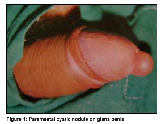
Indian Journal of Dermatology, Venereology and Leprology, Vol. 74, No. 4, July-August, 2008, pp. 430 Net Case Parameatal urethral cyst Aggarwal Kamal, Gupta Sanjeev, Jain VijayKumar, Goel Ashish Department of Surgery, Post Graduate Institute of Medical Sciences and Research, Rohtak Code Number: dv08191 Abstract Cyst formation in the parameatal area of the urethra is an uncommon entity. It was first reported in two male cases as recently as 1956 by Thompson and Lantin. Further reports have been rare. Herein, we report a case of a 21 year-old male having a spherical, cystic swelling 1 cm in size at the external urethral meatus. The diagnosis of parameatal urethral cyst was made and the cyst was excised. Histopathological examination revealed a monolocular cyst lined with transitional cells. The postoperative period was uneventful.Keywords: Male, Parameatal urethral cyst, Penis Case Report A 21 year-old male presented with a cystic swelling of the urethral meatus. The lesion had been present for an unknown number of years, very slowly increasing in size to become more prominent. There were no urinary symptoms, polyuria or stream distortion. The patient′s medical history and a review of all systems indicated general good health. Dermatological examination revealed a spherical cystic swelling, 1 cm in diameter at the external urethral meatus [Figure - 1]. It had a smooth and glistening lining and was fully covered with mucosa. A full urological examination including urography and cysto-urethroscopy was carried out and revealed normal findings. The cyst was completely excised. Histological examination showed a monolocular cyst lined with transitional cells and partly columnar cells with no evidence of inflammation. A diagnosis of parameatal urethral cyst was made. Postoperative recovery was uneventful and a three months′ follow-up period revealed no recurrence. Discussion The parameatal urethral cyst was first reported by Thompson and Lantin in 1956 [1] and about 40 cases have been published since then. The pathogenesis of the cyst is not completely understood. [6] Thompson and Lantin [1] stated that parameatal urethral cysts occurred in the process of delamination or separation of the foreskin from the glans while. Shiraki [7] believed that occlusion of a paraurethral duct was the cause. Oka et al . [8] and Yoshida et al . [9] supported this view while Hill and Ashken pointed out that infection could be a possible cause of the obstruction. The cysts are usually small, averaging about 1 cm in diameter. They occur on the lateral margin of the urethral meatus and may be bilateral. [1] They may be diagnosed as a coincidental finding and may be asymptomatic. However, sometimes they may cause urinary retention, [5] pain during micturition and sexual intercourse, poor cosmesis of the genitalia, and distortion of the urinary stream. When the cyst is traumatized, it may bleed, rupture or become infected. The duration of its occurrence ranges from 16 weeks to two years. The cyst wall epithelium may be columnar, squamous or transitional. The differential diagnosis rests between inflammatory conditions of the urethral meatus, prolapsing ureteroceles (especially in females) and duplications. A full urological examination including urography and cysto-urethroscopy is therefore, advisable. The treatment of choice is complete excision. Simple de-capping results in recurrence. Most of the cases of parameatal urethral cysts reported are in males, but a few female cases have also been reported. [5] A parameatal urethral cyst is a very rare, benign entity; most of the cases reported are in the Japanese population [3] . Extensive literature searches have failed to reveal a single case from the Asian subcontinent. To the best of our knowledge, this is the very first case report from the Asian population. Physicians dealing with sexually transmitted infections must be aware of this condition, not only because of its benign and nonsexually transmitted nature, but also for its timely management.[10] References
Copyright 2008 - Indian Journal of Dermatology, Venereology and Leprology The following images related to this document are available:Photo images[dv08191t2.jpg] [dv08191t1.jpg] [dv08191f1.jpg] |
| |||||||||
