 |
| About Bioline | All Journals | Testimonials | Membership | News |
|
||||||
|
||||||
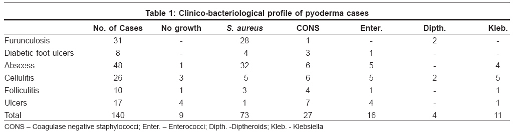
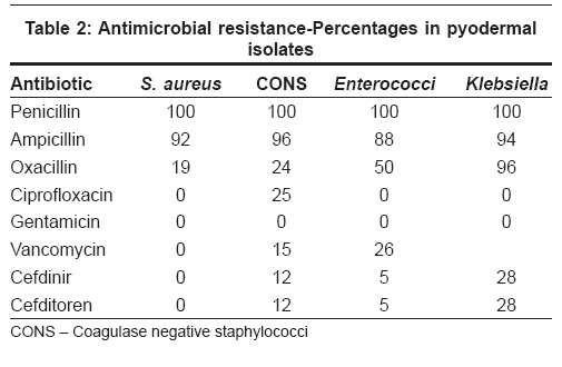
Indian Journal of Dermatology, Venereology and Leprology, Vol. 74, No. 4, July-August, 2008, pp. 430 Net letter In-vitro activities of current antimicrobial agents against isolates of pyoderma Ramana KV, Mohanty SK, Kumar Arun Department of Microbiology, Kamineni Institute of Medical Sciences, Narketpally, Nalgonda-508 254 Code Number: dv08192 Sir, Pyoderma is a common health problem characterized by pyogenic infection of the skin and its appendages. Though easily treatable, the condition is known for its chronicity, recurrence, and other complications. Therefore, timely recognition and prompt bacterial diagnosis with antimicrobial sensitivity is imperative for the effective management and treatment of pyoderma. These lesions are usually produced by Gram positive bacteria, which constitute the majority of cases and less commonly, by Gram negative organisms. [1] The rapid emergence of multidrug resistance in most of the Gram positive bacterial isolates complicates the management of pyoderma and demonstrates the need for more judicious use of antimicrobial agents. [2] The present study has been designed to isolate and identify the bacteria causing pyoderma and their antimicrobial susceptibility patterns to different antibiotics with special reference to the newer generation of cephalosporins-Cefdinir and Cefditoren. This study included 140 pyoderma patients who were attending the Skin OPD of KIMS, Narketpally, between February, 2006 and February, 2007. Cases included in the study had abscess (48), furunculosis (31), cellulitis (26), ulcers (17), folliculitis (10), and diabetic feet (8). A pro-forma consisting of detailed history was taken; clinical and routine investigations were simultaneously done. Pus was collected on sterile cotton swabs from the sites of the lesions and inoculated into Blood Agar and MacConkey′s agar. After aerobic incubation at 37° C, morphology of the isolated organisms was studied and identification was done by standard bacteriological methods. [3] Antimicrobial susceptibility testing of the isolated bacteria against Penicillin (10 µg), Ampicillin (10 µg), Oxacillin (1 µg), Gentamicin (10 µg), Ciprofloxacin (5 µg), Vancomycin (30 µg), Cefdinir (5 µg), and Cefditoren (5 µg) was done by using the disk diffusion method. [4] Of the 140 pyoderma cases studied, males (75, 53%) constituted the majority (females 65, 46%) and most of the cases (64%) were > 40 years old. Staphylococcus aureus (52.1%) was the most commonly isolated organism followed by coagulase negative staphylococci (CONS) (19.2%), e nterococci ( 11.4%), Klebsiella (7.8%) and diphtheroids (2.8%). Nine (6.4%) swabs were culture-negative. Significant findings in the present study include the absence of Streptococci (except Enterococci ). The clinico-bacteriological profile of pyoderma cases in this study is shown in [Table - 1]. The antimicrobial susceptibility testing of isolates revealed greater resistance against penicillin (100%) and ampicillin (92%). Variable resistance against oxacillin was exhibited by S. aureus (19.1%), Enterococci (50%) and CONS (24%). Coagulase-negative S. aureus exhibited a considerable resistance against ciprofloxacin (25%) and vancomycin (15%). All S aureus isolates were sensitive to vancomycin except Enterococci (26%) and CONS (15%). Gentamicin was the only antimicrobial against which all isolates showed 100% sensitivity. S. aureus showed 100% sensitivity to Cefdinir and Cefditoren whereas CONS, Enterococci and Klebsiella showed decreased sensitivities of 88, 95 and 72% respectively, as shown in [Table - 2]. Pyoderma that extends over several months or years is a vexing clinical problem that has not been adequately solved. The results of the present study reveal that S. aureus , CONS, Enterococci -all Gram positive bacteria constitute 85.7% of the causative organisms in pyoderma cases in this region of study. Significant findings of the present study were the absence of Streptococci (except Enterococci ) and the isolation of both coagulase-positive and coagulase-negative Staphylococci in the majority of cases as compared to a study done by Nagmoti et al . who reported Streptococcus pyogenes in 35% of their cases. [5] In the present study, all S. aureus isolates were sensitive to vancomycin and ciprofloxacin. On the other hand, Nagmoti et al . reported resistance to ciprofloxacin in 15% of the S. aureus isolates in their study. [5] In our study, CONS exhibited resistance against ciprofloxacin (25%) and vancomycin (15%) whereas no resistance was seen in a study done by Shoba et al . Pinnaa et al . showed CONS resistance of 9.5 and 2.3% against vancomycin and ciprofloxacin, respectively. [6] Oxacillin resistance of S aureus (19.1%) was considerably low in our study as compared to a study done by Onanuga et al . who reported a resistance of 71.7%. [7] S. aureus (52.1%) is the most common cause of infection as observed in other studies. Resistance against ciprofloxacin (26%), a useful alternative in the treatment of Enterococcal infections, was significantly higher in our study as compared to the findings of Schaberg et al . who reported 15% resistance. [8] Our study′s results suggest that the era of antibiotics has ushered in an unprecedented predominance of Staphylococcal rather than Streptococcal infections. Increasing incidence of methicillin, ciprofloxacin and vancomycin resistance in Staphylococci and Enterococci has limited treatment options. Multidrug-resistant strains also possess the properties of transmissibility and virulence. More recently, possibly as a result of the introduction of newer antimicrobials and their extensive use, strains have been encountered that are resistant to greater numbers of antibiotics. In view of the already existing multidrug-resistant strains, physicians have sought to establish the efficacy of antimicrobial agents against such isolates. References
Copyright 2008 - Indian Journal of Dermatology, Venereology and Leprology The following images related to this document are available:Photo images[dv08192t2.jpg] [dv08192t1.jpg] |
| |||||||||
