 |
| About Bioline | All Journals | Testimonials | Membership | News |
|
||||||
|
||||||
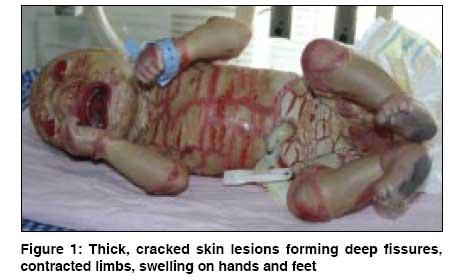
Indian Journal of Dermatology, Venereology and Leprology, Vol. 74, No. 5, September-October, 2008, pp. 487-489 Case Report Neu-Laxova syndrome in an appropriate for gestational age newborn Dilli Dilek, Yasar Handan, Dilmen Ugur, Ceylaner Gulay Department of Pediatrics, Ministry of Health Ankara Training and Research Hospital, Ankara Code Number: dv08207 Abstract Neu-Laxova syndrome is a rare lethal congenital disorder involving multiple systems. Intrauterine growth retardation, ichthyosis, microcephaly, abnormal facial findings, and limb contractures are its key features. We present a case of Neu-Laxova syndrome in a male appropriate for gestational age (AGA) newborn with characteristic features including ichthyosis, microcephaly, severe ectropion, rudimentary ears, eclabion, limb contractures, and hypoplastic genitalia. The patient was born at 38 weeks of gestation to consanguinous Turkish parents. The mother was a 20-year-old primi gravida with lack of prenatal follow-up. Therefore, the case was diagnosed postnatally, and he died 5 days later. Because of the autosomal recessive inheritance of Neu-Laxova syndrome, in countries with high rates of consanguineous marriage, such as Turkey, physicians have to know this syndrome, and serial prenatal ultrasound examinations with genetic counseling should be performed on pregnant women at high risk. To the best of our knowledge, this is the first case described in an AGA newborn.Keywords: Appropriate for gestational age, Newborn, Neu-laxova syndrome Introduction Neu-Laxova syndrome (NLS) is a lethal, autosomal recessive syndrome including multiple congenital abnormalities. This syndrome was first described in three siblings by Neu et al. in 1971. [1] To date, in the medical literature, researchers have reported that NLS was associated with intrauterine growth retardation. Here, we present a case of this rare syndrome diagnosed in an AGA newborn.Case Report A 20-year-old primi gravida woman delivered vaginally a male infant weighing 3000 g (25-50 th centile) and measuring 51 cm (25-50 th centile) in length at 38 weeks of pregnancy. The mother had not been followed up by a gynecologist with prenatal ultrasound examination. Her past medical history was unremarkable except consanguinity (first cousin). At birth, the newborn had a 5-minute APGAR score of < 7 with respiratory distress. His cry was normal, but he was unable to suck effectively. The skin was thickened and covered by edematous, armor-like pale yellowish layer. This layer was cracked, forming deep fissures along the whole body. The neck was short, thick, and edematous. There was severe eclabion with thick and widely separated lips, leaving the mouth open. The chin was underdeveloped. The nasal bridge was flat and the nostrils were patent. The eyes were exophthalmic, and there was severe ectropion. Ears were hypoplastic and deformed. The head circumference was below the 3 rd centile (32 cm) with sloping forehead, completely closed bone sutures, and fontanelles. No scalp hair was present and nails were rudimentary. There was no cleft lip or cleft palate abnormality. There was excessive edema on hands and feet. Limbs were short and contracted. There was no obvious anal opening. External genitalia were hypoplastic, and the testes were not palpable [Figure - 1]. A complete blood count; liver and kidney function tests and serum levels of electrolytes were all normal. Computerized cranial tomography imaging showed lissencephaly and agenesis of the corpus callosum. In addition, chromosome analysis performed on a peripheral blood sample showed a normal karyotype (46, XY). Skin biopsy findings were compatible with ichthyosis. There was extensive hyperkeratosis without parakeratosis. Stratum corneum was thickened compared to stratum malpighi, granular layer was thin, and the basal layer was unremarkable. There was marked papillomatosis and increased fatty tissue beneath the epidermis. Immediately after transfer to our neonatal intensive care unit, the baby was nursed in a humidified incubator maintained at 33°C. Oxygen was administered using a hood. As peripheral venous access was difficult, an umbilical venous line was set up. An extra 25% allowance was provided for fluid and calorie requirements from the first day. After taking appropriate cultures, antibiotics were commenced in order to prevent infection. Vaseline containing lactic acid 5% and local antiseptics were applied topically. Ectropion was covered with eye pads soaked in saline. The plate-like scales split and peeled off, revealing glazed and erythematous skin underneath. He had a cardiorespiratory arrest on the fifth day. No microorganisms grew in the cultures. Discussion In reported cases, NLS has been described as a complex syndrome characterized by marked intrauterine growth restriction (IUGR), ichthyosis, microcephaly, central nervous system (CNS) anomalies and abnormal facial features. The pathologic abnormalities appear to be a result of neuro-ectodermal dysplasia, as evidenced by lack of brain development, eye abnormality, absence of hair, and hyperkeratosis. In our patient, we observed similar findings, including severe ichthyosis, CNS anomalies, microcephaly, and characteristic facial features. Additionally, he had genital abnormalities, including hypoplastic genitalia and bilateral cryptorchidism. In the medical literature, most of the reported cases were associated with IUGR, which was defined as a characteristic feature of the syndrome. [2],[3],[4],[5],[6] However, we would like to draw attention that our case of NLS was diagnosed in an AGA newborn; therefore, we suggest that underlying pathogenesis of IUGR in NLS must be enlightened in further studies. Chromosomal analysis had revealed a normal karyotype and an autosomal recessive inheritance. [3],[4],[5],[6],[7] Russo et al. described a case of Neu-Laxova syndrome in a stillborn female. She was born at 41 weeks of gestation to consanguinous Italian parents. In this case, the affected baby′s chromosomal analysis was normal and parents were first cousins supporting autosomal recessive mode of inheritance. [4] The parents may be offered termination of the pregnancy when the fetus is suspected of NLS. Prenatal ultrasound findings of microcephaly, sloping forehead, exophthalmos, small thorax and abdomen, hypoplastic lungs, syndactyly, hyperextended knees, polyhydramnios, small placenta, and intrauterine growth restriction should indicate NLS. Absence of breathing movements, sucking, swallowing, or normal isolated arm and leg movements may also be detected in the third trimester. [4] Manning et al. presented two patients with NLS with striking prenatal diagnostic findings. Data from these patients had suggested that the NLS represents a heterogeneous phenotype with prenatal ultrasound findings of marked ocular proptosis in a growth-restricted, edematous fetus. [6] In another report from Turkey, Mihci et al. described a case of Neu-Laxova syndrome in a fetus at 22 weeks with ultrasonographic findings of characteristic facial features, limb contractures, kyphosis, and polyhydramnios. [7] Forty-two cases of Neu-Laxova syndrome had been reported by Driggers et al. Only 4 of those cases had been diagnosed prenatally. At 19 weeks of gestation, the authors had presented the earliest reported prenatal diagnosis of Neu-Laxova syndrome in a primi gravida with a non-informative family history. [8] In our case, the mother had a lack of prenatal care and had not undergone any prenatal ultrasonographic examination. Therefore, the patient could not be diagnosed prenatally. Approximately 50% of cases of NLS have been associated with ichthyosis. The inherited ichthyoses are a group of diseases characterized by massive hyperproliferation of the epidermis; and they result in a restrictive, parchment-like encasement of thickened skin and are often associated with genetic syndromes. An extreme sample of these is harlequin fetus, in which the armor-like covering of skin is so restrictive as to be incompatible with life. [9] In our case, NLS was diagnosed due to presence of additional findings such as microcephaly, facial abnormalities, short neck, limb contractures, etc. Because of the autosomal recessive inheritance of NLS, in countries with high rates of consanguinous marriage, such as Turkey, it is important that physicians should consider this syndrome, and serial prenatal ultrasound examinations should be performed on pregnant women at high risk. Moreover, the association between intrauterine growth retardation and NLS is not consistent. References
Copyright 2008 - Indian Journal of Dermatology, Venereology and Leprology The following images related to this document are available:Photo images[dv08207f1.jpg] |
| |||||||||
