 |
| About Bioline | All Journals | Testimonials | Membership | News |
|
||||||
|
||||||
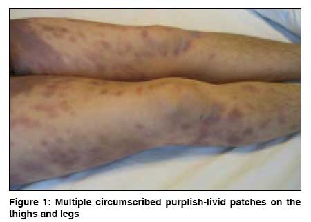
Indian Journal of Dermatology, Venereology and Leprology, Vol. 74, No. 5, September-October, 2008, pp. 511-512 Letter To Editor Ciprofloxacin-induced generalized bullous fixed drug eruption Ada Simin, Yilmaz Sema Department of Dermatology, Baskent University Faculty of Medicine, Ankara Code Number: dv08224 Sir, Ciprofloxacin, a widely used quinolone antibiotic, induces cutaneous adverse drug reactions in about 1% to 2% of treated patients. [1] Urticaria, angioedema, maculopapular exanthem, and photosensitivity are the most frequently documented cutaneous adverse reactions. [2] Only a few cases have been reported in which ciprofloxacin has been implicated in fixed drug eruption (FDE) [3],[4] or more severe drug reactions such as Stevens-Johnson syndrome (SJS) [5] or toxic epidermal necrolysis (TEN). [6] We describe a case of generalized bullous FDE induced by ciprofloxacin. A 57-year-old woman presented with a 3-day history of widespread tender, pigmented patches; and scattered blisters all over her body. The patient stated that these lesions had appeared within a few hours of taking a single dose of oral ciprofloxacin for a respiratory tract infection. She had a previous history of a similar but a more localized reaction after taking the same drug 6 months earlier. She noticed a few residual pigmented macules after that. Her medical history was significant for chronic renal failure, for which she had been on hemodialysis treatment for 5 years. She had been taking perindopril and indapamide for hypertension and congestive heart failure during the same period. The patient denied taking any other new drug in the preceding days. Physical examination revealed extensive purplish-livid patches covering almost 60% of the total body surface. Multiple well-circumscribed patches were observed on the arms and legs [Figure - 1]. Some of the patches were studded with flaccid vesicles and bullae over the buttocks. Eroded areas were also noted on the arms and legs. Pseudo-Nikolsky′s sign was positive on some of the purplish-livid patches. The mucous membranes, the palms and soles, and the face were not involved. Her temperature was 37°C, and her other vital signs were within normal range. The patient was hospitalized with the differential diagnosis of generalized bullous FDE, SJS, and TEN. Histopathological examination of a punch biopsy specimen taken from a flaccid bulla overlying the large purplish-livid patch on the left buttock showed necrosis of epidermal keratinocytes with subepidermal clefting . Perivascular mixed inflammatory infiltrate containing eosinophils and neutrophils, and prominent pigmentary incontinence were also observed within the dermis. Direct immunofluorescence revealed no IgG and C3 deposition at the basement membrane zone. Laboratory investigations showed the following values: C-reactive protein of 65 mg/L (normal range, < 10 mg/L); white blood cell count of 20.5x10 9 /L (normal range, 4.5-11x10 9 /L) with 70% neutrophils (normal range, 40%-72%), hemoglobin 8.2 g/dL (normal range, 12-16 g/dL), and blood urea nitrogen 65 mg/dL (normal range, 6-21 mg/dL); and a creatinine level of 6.2 mg/dL (normal range, 0.5-1.3 mg/dL). A chest x-ray revealed increased opacities in the middle and lower areas, indicating bilateral pneumonic infiltrates. The patient was diagnosed as having ciprofloxacin-induced generalized bullous FDE. We initiated oral prednisolone (1 mg/kg/d) therapy. She was also given oral clarithromycin (1 g/d) treatment for suspicion of atypical pneumonia. The vesicles, bullae, and eroded areas disappeared after 1 week, leaving a dusky-brown residual hyperpigmentation. Oral prednisolone was discontinued after 10 days. Understandably, the patient refused oral or topical provocation testing. The severity of reactions in FDE may increase after repeated exposures to the drug and very rarely progress to a clinical state - so-called generalized bullous FDE. Generalized bullous FDE with its characteristic multiple large, purplish-livid patches, at times with flaccid blisters, may be clinically misdiagnosed as SJS or TEN. In generalized bullous FDE, the disease onset is within hours after the drug exposure. A diagnostic hallmark is the reappearance of the lesions over the previously affected sites, when the offending drug is reused. The extent of epidermal detachment has not been described in previous case reports of generalized bullous FDE. Mucosal sites are usually spared, and constitutional symptoms are mild. Also, the lesions heal rapidly without complication, leaving residual hyperpigmentation. Histopathological differentiation of generalized bullous FDE from SJS and TEN may be challenging. Epidermal changes varying from a few scattered necrotic keratinocytes to full-thickness epidermal necrosis cannot be distinguished in all 3 conditions. In FDE, a mixed inflammatory infiltrate containing not only lymphocytes but also neutrophils and eosinophils is present around both the superficial and deep plexus. In SJS and TEN, the infiltrate is mainly lymphohistiocytic and tends to be located around the superficial plexus. [7] In our patient, the temporal correlation with the drug, previous history of a milder reaction, lack of involvement of mucosal sites, and the rapid and uneventful recovery that left residual hyperpigmentation led us to consider a diagnosis of generalized bullous FDE due to ciprofloxacin. The histopathological findings were also consistent with this diagnosis. Perhaps only a provocation test would provide sufficient proof. However, taking into account the severity of the lesions, we considered it unethical to perform an oral provocation test. Furthermore, the value of topical provocation with patch testing and intracutaneous testing in ciprofloxacin-induced fixed drug eruption is not well established. The present case documents that ciprofloxacin may cause generalized bullous FDE. References
Copyright 2008 - Indian Journal of Dermatology, Venereology and Leprology The following images related to this document are available:Photo images[dv08224f1.jpg] |
| |||||||||
