 |
| About Bioline | All Journals | Testimonials | Membership | News |
|
||||||
|
||||||
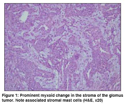
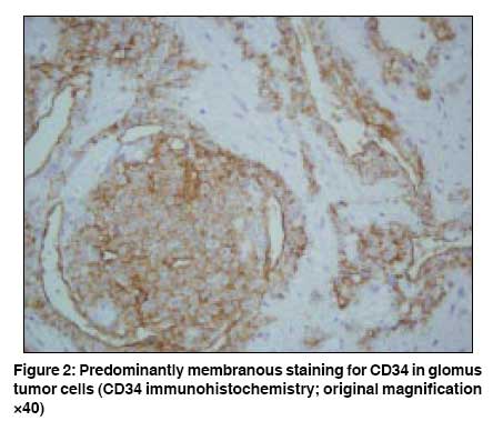
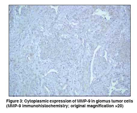
Indian Journal of Dermatology, Venereology and Leprology, Vol. 74, No. 5, September-October, 2008, pp. 519-521 Letter To Editor Matrix metalloproteinase-9 expression in a CD34-positive glomus tumor with myxoid stromal change Pabuccuoglu Ugur, Lebe Banu Dokuz Eylül University, School of Medicine, Department of Pathology, Inciralti/Izmir Code Number: dv08230 Sir, Glomus tumors commonly present as solitary lesions and typically occur in the dermis or subcutis of extremities of the adults. Subungual region of the finger is the most common localization for these tumors. Microscopically, a glomus tumor has distinctive histological features. It is a well-demarcated or encapsulated dermal or subcutaneous lesion composed of sheets or nests of uniform glomus cells surrounding blood vessels. [1] Immunohistochemically, glomus tumor cells express vimentin, actin, and myosin, while desmin is generally negative. [2] A 56-year-old woman was admitted with a complaint of a nodule located at the lateral side of the fifth finger of her right hand. The lesion was completely excised and submitted for histopathological examination. On gross pathology, a nodular encapsulated lesion measuring 8x5 mm was observed at the cut surface of the excised specimen. At histopathological examination, a well-demarcated tumoral lesion with a fibrous capsule was noted in the reticular dermis. Tumor tissue was composed of nests of rather uniform cells with acidophilic cytoplasm and round-to-oval nuclei and these nests were interrupted by blood vessels. Tumor cells surrounded the blood vessels in many areas. A prominent myxoid change was noted in the tumor stroma [Figure - 1]. Tryptase immunohistochemistry (1/100, Neomarkers, Fremont CA) highlighted the mast cells scattered around. The number of mast cells was over 20 per high-power field (HPF). No diffuse cellular atypia or mitotic activity was noted within the tumor tissue. Immunohistochemically, smooth muscle actin (1/200, Neomarkers, Fremont CA) and CD34 (1/50, Neomarkers, Fremont, CA) were positive in all tumor cells [Figure - 2]. Endothelial cells also showed diffuse staining for CD34. The histopathologic diagnosis was that of a CD34-positive glomus tumor with a myxoid stroma. Regarding the myxoid stromal change encountered in the present case, additional immunohistochemical staining was performed for matrix metalloproteinase-2 (MMP-2) (pre-diluted, Neomarkers, Fremont, CA) and matrix metalloproteinase-9 (MMP-9) (1/25, Neomarkers, Fremont, CA) in order to find out if they are expressed within the tumor tissue. MMP-9 immunohistochemistry showed a consistent but rather weak cytoplasmic staining in most of the tumor cells [Figure - 3]. Mast cell cytoplasm was also similarly stained. Stromal cells and endothelium were negative for MMP-9. MMP-2 was found to be negative in all cellular components throughout the lesion. The stroma of glomus tumors is generally fibrous. Prominent stromal myxoid degeneration is an uncommon finding. [3],[4],[5] Mentzel et al, [3] reported 6 cases of glomus tumors showing myxoid stromal changes and associated co-expression of actin and CD34 by the tumor cells. The authors also noted that CD34 expression by tumor cells was limited to glomus tumors with myxoid stroma. Interestingly, in the present case, co-expression of actin and CD34 was also associated with MMP-9 expression by the tumor cells. MMPs are a family of proteolytic enzymes involved in the degradation of many constituents of basement membrane and extracellular matrix, and they are known to play a role in the tumor invasion and metastasis. MMP-9 is also known as 92 kD gelatinase (gelatinase-B). [6] Hence it may be speculated that the myxoid stromal change in the present case may be related to MMP-9 expression by the glomus tumor cells. Hisa et al, [4] have reported a correlation between the number of glomus cells and the extent of mucinous degeneration. However, mast cells are also known to produce MMP-9 to mediate extracellular matrix degradation. [7] Regarding this mast cell function, it is also reasonable to assume that MMP-9 released by mast cells might be responsible for the myxoid stromal change encountered in some glomus tumors. Nevertheless, the presence of variable numbers of mast cells is a usual feature for these tumors. Daugaard et al, [2] graded the number of mast cells in 18 glomus tumors and found that 3 had many (more than 20 per HPF) stromal mast cells, whereas 14 had few (less than 10 per HPF). In their study, no myxoid change was reported in glomus tumor cases having a large number of mast cells. Though an increase in the mast cell population was noted in the present case, myxoid degeneration cannot be solely attributed to it since MMP-9 was consistently expressed in glomus tumor cells as well. Regarding these findings, contribution of both cells to myxoid degeneration is likely. In conclusion, besides the co-expression of actin and CD34, we also noted MMP-9 expression by the tumor cells in a glomus tumor with myxoid stroma. It may be assumed that immunophenotypic change of tumor cells to express MMP-9, as well as an increased number of MMP-9-expressing mast cells, may be responsible for this myxoid stromal change in the present case. Additional studies on MMP expression and mast cell quantification in CD34-positive myxoid glomus tumors may highlight this issue. References
Copyright 2008 - Indian Journal of Dermatology, Venereology and Leprology The following images related to this document are available:Photo images[dv08230f2.jpg] [dv08230f1.jpg] [dv08230f3.jpg] |
| |||||||||
