 |
| About Bioline | All Journals | Testimonials | Membership | News |
|
||||||
|
||||||
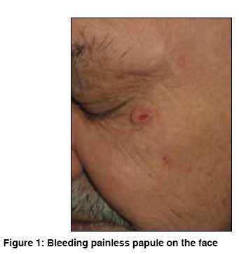
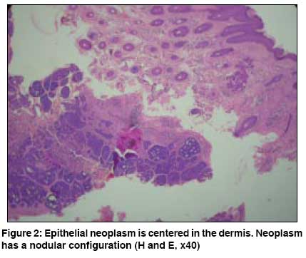
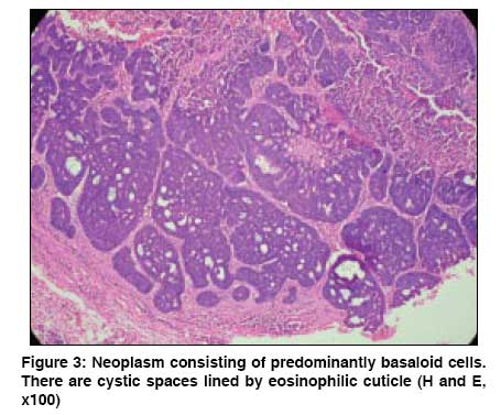
Indian Journal of Dermatology, Venereology and Leprology, Vol. 74, No. 5, September-October, 2008, pp. 541-542 Quiz A painless punctured papule Verma Shyam Consultant Dermatologist, Vadodara, Gujrat Code Number: dv08236 A 73-year-old building contractor presented to this clinic with a bleeding lesion on the face. The lesion was present since 8 months and there were 3 episodes of scanty bleeding in the past. On examination there was a skin-colored firm, non-tender, freely movable, and well-defined papule inferior to the lateral canthus of the left eye [Figure - 1]. It had visibly undergone a puncture and looked as if it had been physically scooped out. Pressure did not express any fluid or solid material from it. There was no similar lesion elsewhere on the body. No regional nodes were palpable. Findings from an excisional biopsy are show in [Figure - 2] and [Figure - 3]. What is your Diagnosis ? Diagnosis: Eccrine spiradenoma The excisional biopsy showed an epithelial neoplasm in mid-dermis that was extremely well circumscribed. The neoplastic cells were basaloid and were arranged in closely crowded reticulated pattern that formed variously sized nodules. Within these nodules, several scattered ductal structures lined by eosinophilic cuticle were seen. Several darker lymphocyte-like cells dotted the neoplastic nodules. Surrounding collagen was slightly denser. Discussion Eccrine spiradenoma, also known as spiradenoma, is an uncommon well-differentiated benign tumor originating from the sweat gland. [1],[2] It is best described as a firm dermal skin colored or bluish nodule and is usually solitary; however, occasionally multiple lesions may be present, and some occurring in linear or segmental pattern have been documented. [3],[4] It is usually found on the ventral aspect of trunk and proximal limbs. Histopathologically, eccrine spiradenoma consists of one or more large, sharply defined basophilic nodules in the dermis, also described as ′cannon balls′ or ′big blue balls′ in the dermis. [5] They are not attached to the overlying epidermis and sometimes extend into the subcutis. They consist of groups of cells in cords or islands or sheets. Two types of cells are found in these nodules. There are small, dark, basaloid cells with hyperchromatic nuclei; and cells with large, pale vesicular and ovoid nuclei. Pale cells tend to be at the center of the lesions. [5] Familial cases have been described. [6] When painful, it can lead to a misdiagnosis of leiomyoma, glomus tumor, neuroma, or angiolipoma. Malignant transformation is uncommon but is documented especially in longstanding cases. [7],[8] When it occurs, the rate of metastasis can be as high as 50% and can lead to death. Treatment is an adequate surgical excision as it may recur if not removed completely. This case was unusual in its clinical morphology and it bled repeatedly. References
Copyright 2008 - Indian Journal of Dermatology, Venereology and Leprology The following images related to this document are available:Photo images[dv08236f3.jpg] [dv08236f2.jpg] [dv08236f1.jpg] |
| |||||||||
