 |
| About Bioline | All Journals | Testimonials | Membership | News |
|
||||||
|
||||||
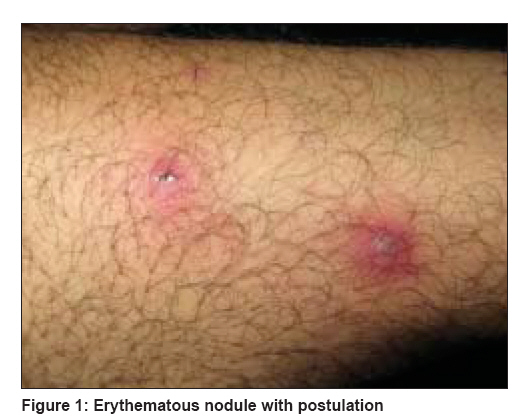
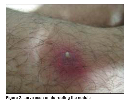
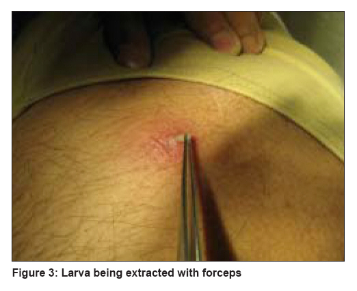
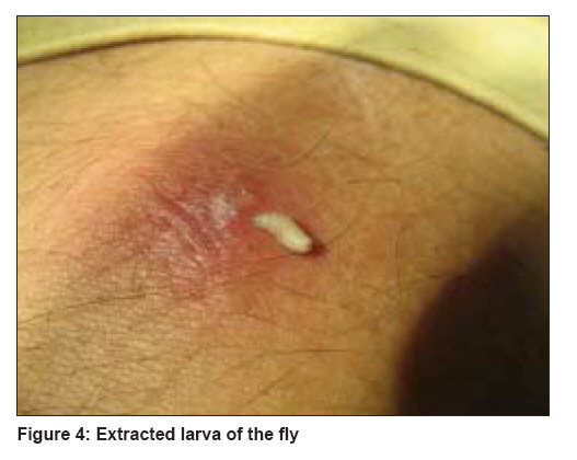
Indian Journal of Dermatology, Venereology and Leprology, Vol. 74, No. 6, November-December, 2008, pp. 679-681 Letter To Editor Furuncular myiasis mimicking pyoderma Sharma Parikshit, Pai HarshaS, Pai GaneshS Department of Skin and V.D., Kasturba Medical College, Mangalore (Karnataka) Code Number: dv08282 Sir, Myiasis is the term applied to the infestation of live humans and vertebrate animals with the larva (maggots) of Diptera (two-winged) flies. [1],[2] Furuncular myiasis as a result of Cordylobia anthropophaga infestation has been endemic in West African sub-region for more than 135 years. [3] The other flies that cause furuncular myiasis include Cordylobia rhodaini (Lund fly, found in the rain forest of Africa) and Dermatobia hominis (human botfly).[3] The mode of transmission of Dermatobia hominis differs from that of Cordylobia species. The eggs of D. hominis are carried to the host by bloodsucking insects such as mosquito and tics, and the hatched larvae invade exposed skin of trunks, head, and limbs. The eggs of Cordylobia species are, however, deposited on the soiled or wet and soiled clothes hung outside for drying. The hatched larvae invade unexposed skin (of the buttocks, trunk, limbs, and penis) in contact with the wet clothes. A 45-year-old man, resident of Lagos, Nigeria, presented with a 3-day history of multiple painful lesions over the limbs and trunk incubated during travel to India. He gave a history of wearing washed clothes that were hung out to dry and not ironed. He also complained of some movement felt within some of the lesions. Examination revealed multiple, tender, erythematous nodules with central pustulation [Figure - 1]. Lesions were present on the thigh, legs, and the prepuce. On deroofing the nodule, a larva was seen and gently extracted [Figure - 2]. As the larva is known to infest the eyes, a detailed ophthalmological examination of the anterior and posterior chambers was done, which revealed no abnormality. The diagnosis of cutaneous myiasis was made. Eight larvae were extracted from the affected lesions. They were identified as the larvae of Cordylobia anthropophaga by the parasitologist. Liquid nitrogen was sprayed on each lesion before attempting extraction [Figure - 3],[Figure - 4]. This served a dual purpose - stiffening the larva that facilitated extraction and also anesthetizing the skin. Further a 7-day course of amoxycillin-clavulanic acid and metronidazole was given to prevent any secondary infection. Diagnosis of furuncular myiasis is mainly clinical. Main diagnostic features are recent travel to endemic area, one or more non-healing lesions on the skin, symptoms of pruritus, movement under skin, or pain. Other features include serous or serosanguinous discharge from a central punctum and a small white threadlike structure protruding from the lesion. The diagnosis is confirmed by extraction of the larvae. The goal of treatment is to remove the larva and treat any associated infection with antibiotics. The lesions heal rapidly after larva is removed. Traditional methods of removing larva include occluding the punctum with pork fat, mineral oil, petrolatum, or butter, thus suffocating the larva, which forces it to wriggle out. [4] Surgical removal is done by enlarging the punctum by cruciate incision, and this enables removal of intact larva. [5] Forceps may also be used. This must be done carefully and the entire larva extracted, as any remnant may provoke an inflammatory response. Cutaneous myiasis is an uncommon condition. In this era of international travels, an immigrant may get infested with the larva and present with lesions, which in a non-endemic area like India can be misdiagnosed as a simple pyoderma. Hence an awareness of its clinical features is essential to avoid unnecessary delay in the diagnosis and the treatment. General improvement of sanitation, personal hygiene, and exterminating the flies by insecticide are helpful in prevention. Simple measures such as washing clothes thoroughly and drying and ironing of clothes are also necessary to reduce the risk of this human myiasis. References
Copyright 2008 - Indian Journal of Dermatology, Venereology and Leprology The following images related to this document are available:Photo images[dv08282f3.jpg] [dv08282f4.jpg] [dv08282f2.jpg] [dv08282f1.jpg] |
| |||||||||
