 |
| About Bioline | All Journals | Testimonials | Membership | News |
|
||||||
|
||||||
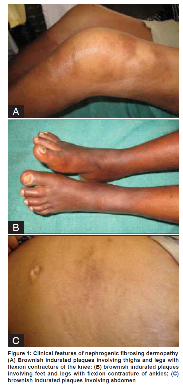
Indian Journal of Dermatology, Venereology and Leprology, Vol. 75, No. 1, January-February, 2009, pp. 63-67 Case Report Nephrogenic fibrosing dermopathy Ragunatha S, Palit Aparna, Inamadar ArunC, Madraki RavindraM, Yelikar BR Department of Dermatology, Venereology and Leprosy, BLDEA's SBMP Medical College, Hospital and Research Centre, Bijapur - 586 103, Karnataka Code Number: dv09015 Abstract An adult female patient on hemodialysis for chronic renal failure presented with large, brownish, and indurated plaques with bound-down skin on both lower limbs and abdomen along with difficulty in movement of the legs. Histopathological features revealed thick collagen bundles admixed with mucin and intercalating spindle-like cells characteristic of nephrogenic fibrosing dermopathy (NFD). Immunohistochemical study showed prominent CD68 positivity and weak CD34 positivity suggesting that the plaques were more than 20-weeks old. NFD in patients with chronic renal failure of unknown cause is a poor prognostic indicator. Early detection before the development of contracture and prompt treatment of NFD and underlying renal failure may reverse this disabling condition.Keywords: Nephrogenic fibrosing dermopathy, Nephrogenic systemic fibrosis, Dialysis, Renal failure Introduction Nephrogenic fibrosing dermopathy (NFD) is a recently described fibrosing disorder seen in the setting of renal failure with or without dialysis. The process of fibrosis in patients with NFD has been reported to involve underlying structures and also internal organs. Therefore, some authors prefer to designate the condition as nephrogenic systemic fibrosis. Though many endogenous and exogenous factors have been implicated as potential associated factors, the exact etiology of this disorder remains unclear. The same holds good for the management of disease. We are reporting clinical, histopathological, and immunohistochemical features of NFD which may add to the present understanding of this new disease entity. [1],[2],[3],[4]Case Report A 30-year-old female patient on hemodialysis for chronic renal failure was referred to dermatology clinic for pruritic skin lesions and difficulty in movement of the lower limbs for the past 15 days. The underlying cause for her chronic renal failure was unknown, and she was undergoing hemodialysis for the past 9 months. There was no history of exposure to radiocontrast agents or invasive procedures except for arteriovenous fistula performed for dialysis. The skin lesions were insidious in onset and gradually progressive involving both the lower limbs resulting in flexion contractures. On examination, there were multiple large, irregular, brownish indurated plaques distributed predominantly over thighs, ankles, legs, and abdomen [Figure 1a],[Figure 1b],[Figure 1c]. The overlying skin was shiny. On palpation, the plaques were mildly tender, bound down, and woody hard in consistency. Laboratory investigations revealed a low hemoglobin (4.5 g/dL) and raised erythrocyte sedimentation rate (60 mm after 1 hour), blood urea (72 mg/dl), and serum creatinine (3.6 mg/dl) levels. VDRL test was nonreactive and ANA was negative. The histopathology of skin biopsy specimen revealed thin epidermis, and thickened collagen bundles with surrounding clefts throughout the dermis. Mild inflammatory infiltrate, comprised mainly of lymphocytes and macrophages was present especially in the upper dermis. Spindle-shaped cells were scattered in between the collagen bundles [Figure - 2]. The alcian blue stain showed deposition of mucin in between the collagen bundles [Figure - 3]. On immunohistochemical study, the section showed prominent intracytoplasmic CD68 positivity [Figure - 4] and very weak CD34 positivity [Figure - 5]. With the above clinical and laboratory findings, NFD was diagnosed and the patient was started on oral prednisolone 1 mg/kg/day. After two weeks of treatment, there was mild improvement in the movement of limbs. Later, within a month, patient succumbed to worsened chronic renal failure. Discussion Since its first description in 1997, around 200 cases have been registered in the international NFD registry. [1] The clinical presentation of NFD has been consistent with no reported clinical variants. There is no age, sex, or racial predilection for the development of this disorder. [2] Clinically, it is characterized by mildly pruritic, tender, and browny indurated plaques with woody hard consistency, predominantly distributed over thighs, legs, and trunk, sparing the face. In later stage, there may be restricted joint mobility with subsequent flexion contractures of the limbs. [2],[3] Yellowish scleral plaques may be present in some cases. [4] The fibrosing process is not only restricted to the dermis, but also involves underlying fascia and muscle, breast, myocardium, diaphragm, renal tubules, and rete testes. [1] The presence of CD34+, procollagen-1 positive spindle-like cells, and CD68+ mononuclear cells in between the thickened collagen bundles is characteristic of NFD. Other histopathological features include increased elastic fibers and deposition of variable amount of mucin in the dermis. [5] Abnormal calcification of dermis and subcutaneous tissue has also been reported. [4],[5] The clinical and histopathological differential diagnosis include those disorders with sclerosis, fibroblast proliferation, and mucin deposition, viz. scleromyxedema, morphea, systemic sclerosis, eosinophilic fascitis, and eosinophilia-myalgia syndrome. The characteristic clinical and histopathological features, underlying renal failure, and sparing of face easily distinguish NFD from other sclerosing disorders. [5] Though clinical and histopathological features of NFD are well delineated, the etiopathogenesis is not yet clear. The disorder is common in patients with renal failure, undergoing hemodialysis or peritoneal dialysis. However, it has also been reported in patients who are not on dialysis. But the common denominator in all the patients reported till date, is renal failure. [6] This suggests that the presence of renal failure is a prerequisite for the development of NFD. The involvement of internal organs and presence of circulating fibrocytes (CD34+ cells) in the lesional skin suggest that NFD is a systemic disease. Normally, these circulating fibrocytes induce scar formation by secreting cytokines in the area of tissue injury. Therefore, the fibrosing process that occurs in NFD is probably due to inappropriate recruitment and activation of circulating fibrocytes in a patient with renal failure. [4] In view of the fact that NFD is a recently recognized disease, it has been postulated that the new drugs, chemicals, or radiocontrast agents which were introduced around the same time as the first cases of NFD, are the probable etiological factors. These get deposited in the peripheral tissue and act as allergens to incite the pathogenetic process. [1],[2] Other than gadolinium link, there has been no clear association between NFD and the many correlated drugs, chemicals, or radiocontrast agents. Recently, several studies have demonstrated temporal association between the use of gadolinium and the development of NFD. Gadolinium is a radiocontrast agent that has predominant renal clearance. [6] Godadiamide, a nonionic chelate of gadolinium and diethylene triamine pentaacetic acid bismethylamide, is less stable and undergoes transmetallation with endogenous ions. These poorly soluble ions can form salt precipitates with phosphate and other anions, which are usually elevated in renal failure, resulting in tissue deposition and inflammation. In addition, gadolinium has been detected in the lesional skin of NFD. These deposits may stimulate resident circulating fibrocytes to synthesize extracellular matrix and collagen. [1] Apart from this, other factors also seem to contribute to the pathogenesis. Certain endogenous and exogenous clinical events have been reported to occur frequently prior to the development of NFD. The former includes thrombotic events, hypercoagulable states (antiphospholipid antibodies), hepatic disease (hepatitis C), generalized edema, and the latter includes drugs ( e.g., erythropoietin), hemodialysis or peritoneal dialysis, infections, prior vascular surgeries, and diagnostic procedures. Though common in the setting of renal failure, these may also act as contributing factors by inciting endothelial or tissue injury and consequently recruiting circulating fibrocytes to the site. [1],[6],[7] In addition, the profibrotic cytokines like TGF β and bFGF are frequently elevated in the dialysate of peritoneal dialysis and in hepatitis C patients. Therefore, patients with hepatitis C undergoing hemodialysis are at increased risk of developing NFD. [3] Thus, in a patient with renal failure the probable etiological factors and contributing factors together perpetuate the fibrosing process in the skin, underlying structures, and internal organs [Figure - 6]. In the present case, the patient presented with classical clinical features of NFD. But there were fewer CD34+ cells correlating with the duration of lesions. In lesions of duration of less than 20 weeks, the infiltration is usually dense. However, the same becomes less conspicuous in the lesions of duration more than 20 weeks. [5] Very weak positivity has been reported in a patient with contracture. [6] Depending upon the type of renal failure and course of the disease, NFD has been divided into seven subtypes. According to this classification, the patients with acute renal failure and chronic renal failure of unknown cause are more likely to develop transient and chronic NFD, respectively. [4] The present case belongs to the latter subtype which has a poor prognosis. There is no specific therapy for NFD. The treatment is challenging because the disease may appear and regress spontaneously in relation with renal failure making the evaluation of efficacy of the drug difficult, and sometimes, it may persist even after return of renal functions to normal. Several drugs have been used including, systemic and topical corticosteroids, immunosuppressants, oral retinoids, intravenous immunoglobulins, psoralen-UVA therapy, interferon-a, plasmapheresis, topical calcipotriene, and extracorporeal photopheresis (ECP). The therapeutic effects of many of these drugs have been variable. [1],[4] However, ECP has been reported to be effective in improving some of the symptoms of NFD. This can benefit the patient significantly, at least by improving the quality of life or by arresting the progression of disease. The treatment would be useful if the patients are treated before the development of extensive sclerosis. [1] Since it is a newly recognized entity and less symptomatic at an early stage, patients are likely to consult the physician only when joint contracture develops, as in the present case. Hence, early detection and prompt treatment of NFD, and underlying renal disease may improve the prognosis of the condition. Further studies are required to conclusively ascertain the etiology, pathogenesis, risk factors, and effective therapeutic agents.[Table - 1] References
Copyright 2009 - Indian Journal of Dermatology, Venereology and Leprology The following images related to this document are available:Photo images[dv09015f4.jpg] [dv09015f3.jpg] [dv09015f5.jpg] [dv09015f6.jpg] [dv09015f1.jpg] [dv09015t1.jpg] [dv09015f2.jpg] |
| |||||||||

![[Figure - 2]](/showimage?dv/photo/dv09015f2.jpg)
![[Figure - 3]](/showimage?dv/photo/dv09015f3.jpg)
![[Figure - 4]](/showimage?dv/photo/dv09015f4.jpg)
![[Figure - 5]](/showimage?dv/photo/dv09015f5.jpg)
![[Figure - 6]](/showimage?dv/photo/dv09015f6.jpg)
![[Table - 1]](/showimage?dv/photo/dv09015t1.jpg)