 |
| About Bioline | All Journals | Testimonials | Membership | News |
|
||||||
|
||||||
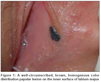
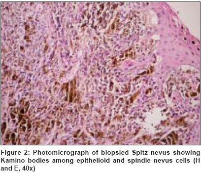
Indian Journal of Dermatology, Venereology and Leprology, Vol. 75, No. 2, March-April, 2009, pp. 167-169 Case Report Spitz nevus of the genital mucosa Polat Mualla, Topcuoglu MehmetAta, Tahtaci Yasemin, Hapa Asli, Yilmaz Fahri Department of Dermatology, Izzet Baysal Medical Faculty, Abant Izzet Baysal University, Bolu Code Number: dv09046 Abstract We herein report an 11-year-old girl who came to our clinic with a swelling on the genital area of 2 months duration. Dermatological examination of the patient was performed and a pigmented lesion was found on the inner surface of the labium majus of the mucosa. The lesion was well circumscribed and approximately 1 cm in diameter, with homogenous color distribution. The patient was diagnosed as Spitz nevus on the basis of clinical and histopathological findings. Our case is probably the first reported case of Spitz nevus localized to the genital mucosa in the English literature.Keywords: Spitz nevus, genital mucosa Introduction Spitz nevus was first described by Sophie Spitz in 1948. [1] Spitz nevus, which is generally seen in children and adolescents, was originally called as "juvenile melanoma" as it is histopathologically similar to malignant melanoma. [2] Although it is mostly seen on the skin of the face, hands, neck, trunk and lower extremities, it is rarely seen on other areas and on the mucosa. [3],[4],[5],[6],[7],[8],[9] Review of the English language literature did not reveal a Spitz nevus located on the genital mucosa. In this report, we report such a finding in our case.Case Report An 11-year-old girl came to our clinic with a swelling on the genital area of 2 months duration. Her history and family′s medical history were unremarkable. On dermatological examination, a pigmented lesion was found on the inner surface of the labium majus of the genitalia. The lesion was well circumscribed, approximately 1 cm in diameter that had homogenous color distribution and was raised by 2 mm from the mucosal surface [Figure - 1]. Clinical examination did not reveal any inguinal lymphadenopathy. Total surgical excision of the lesion was performed. The final diagnosis was Spitz nevus according to clinical and histopatological examinations [Figure - 2]. The patient and her family were informed of the diagnosis and she is currently under periodic follow-up.Discussion Spitz nevus is a benign, asymptomatic, papulonodular lesion ranging from 3 to 10 mm in diameter, which is seen especially in children and adolescents. It usually appears as a pink or light-brown, smooth-surfaced, well-circumscribed, hairless lesion, which is firm on palpation. There can be telangiectasia, erosion or crusts on the surface of the lesion. Even if Spitz nevus occurs as a solitary lesion in most of the cases, it can also be seen as multiple lesions. [8] Although Spitz nevus is usually an acquired condition, there are reports of congenital Spitz nevi in the English literature. [10] Although it is generally seen on the skin of the face, hand, neck, trunk and lower extremities, it can be rarely seen on some other parts of the body. Examples of unusual localizations of Spitz nevus are glans penis and corpus penis. [7],[8],[9] Although extremely rare, Spitz nevus can also be seen on mucosal surfaces. Only three Spitz nevus cases on the oral mucosa have been reported so far. The first one of them, "Spitz nevus of the palate" was reported by the Nikai et al. [3] He reported the first case of Spitz nevus of the oral mucosa. It was in a 4-year-old girl with a congenital non-pigmented nodular lesion on the anterior portion of the palate. [3] Another case was noted in a 24-year-old male patient, which was located on the upper lip mucosa for the last 4 months. [4] Eroπlu et al. diagnosed a 14-year-old girl with Spitz nevus of the tongue on the basis of histopathological and immunohistochemical findings of the lesion. [5] In the English literature, it has been reported that Spitz nevus can be rarely localized on the conjuctiva. [6] In our case, Spitz nevus was seen on the inner surface of the labium majus, which has not been reported so far. Diagnosis of Spitz nevus depends on clinical, dermatoscopic and histopathological findings. As the genital lesion was a pigmented lesion, thus diagnosis in our case of Spitz nevus was unlikely. Outburst star pattern is expected on dermatoscopic examination but, unfortunately, the patient did not co-operate for such an examination. Hence, we skipped this and total surgical excision for definite diagnosis was done. Histological features consistent with a Spitz nevus include mostly large epithelioid and/or spindle-shaped melanocytes with abundant amphophilic cytoplasm and classically are symmetrical, small, contain clefts between nests of melanocytes and keratinocytes, have sharp lateral demarcation, exhibit maturation of melanocytes and contain the eosinophilic globules (Kamino bodies). [10] In the differential diagnosis, malignant melanoma, nevomelanocytic nevus, atypical melanocytic nevus, pyogenic granuloma, hemangioma, juvenile xanthogranuloma, dermatofibroma, isolated mastocytoma and pseudolymphoma should be considered. Despite the benign nature of Spitz nevus, the recommended management is total excision. [4] Local recurrence is seen frequently on incomplete removal. Cases of malignant transformation have been documented and, due to the histological similarities between a Spitz nevus and malignant melanoma, close follow-up is advised. [11] In this report, we present the first case of genital mucosa-localized Spitz nevus. To the best our knowledge, it is the first report with such localization so far. Thus, Spitz nevus should be considered in differential diagnosis of pigmented lesions of genital mucosa. References
Copyright 2009 - Indian Journal of Dermatology, Venereology and Leprology The following images related to this document are available:Photo images[dv09046f2.jpg] [dv09046f1.jpg] |
| |||||||||
