 |
| About Bioline | All Journals | Testimonials | Membership | News |
|
||||||
|
||||||
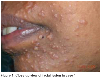
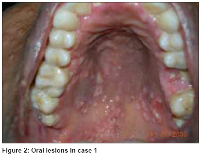
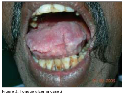
Indian Journal of Dermatology, Venereology and Leprology, Vol. 75, No. 2, March-April, 2009, pp. 173-176 Case Report Two unusual cases of histoplasmosis in human immunodeficiency virus-infected individuals Bhagwat PV, Hanumanthayya K, Tophakhane RS, Rathod RM Department of Skin and STD, Karnataka Institute of Medical Sciences, Hubli - 580 022, Karnataka Code Number: dv09048 Abstract Histoplasmosis, also called as Darling's disease, is caused by the dimorphic fungus, Histoplasma capsulatum. In India, several cases of histoplasmosis have been reported since 1954, but in only a few cases could the diagnosis be confirmed by fungal culture. Disseminated histoplasmosis in adults is often associated with immunosuppression, as in human immunodeficiency virus (HIV) infection. Oral lesions are seen in 30-50% of the patients. Here, we are reporting two histoplasmosis cases, one with disseminated histoplasmosis with extensive skin and oral lesions in a known HIV-positive patient and the second case presenting with ulcer of the tongue, found to be HIV positive on investigation. It is important to consider histoplasmosis as one of the differential diagnosis of oral lesions in HIV-infected individuals.Keywords: Disseminated histoplasmosis, human immunodeficiency virus infection, Oral histoplasmosis Introduction Histoplasmosis is a granulomatous fungal disease caused by Histoplasma capsulatum , which is found in soil rich with excreta of bats and birds. This disease has variable clinical features. Upper aerodigestive lesions are chiefly associated with systemic disease, especially affecting patients with immunosuppression, as in human immunodeficiency virus (HIV) infection. [1] Disseminated disease usually occurs in immunocompromised patients or in patients with chronic illness. Although relatively uncommon, histoplasmosis has been reported in patients with acquired immunodeficiency virus syndrome and oral lesions have varied presentations. [2] In the setting of disseminated disease, oral lesions are present in 30-50% of the patients and may occur in almost every part of the oral mucosa. The most common sites are the tongue, palate and buccal mucosa. In some cases, oral lesions appear to be the primary or only manifestation of the disease. [3],[4] Here, we are reporting two cases of histoplasmosis, one with disseminated disease with lesions on the skin, oral cavity and viscera and the second case, presenting for the first time, with only oral lesions and found to be in an advanced-stage of HIV infection on investigation.Case Reports Case 1 On examination, she had multiple, discrete, pearly white papules and plaques, mildly tender, distributed over the face, neck, abdomen, back, oral cavity and vulva. Some of them showed spontaneous ulceration and crusting. The lesions resembled molluscum contagiosum [Figure - 1] and [Figure - 2]. She had generalized lymphadenopathy with non-tender hepatosplenomegaly. The lymph nodes were discrete, firm, non-tender and mobile. Examination of the respiratory, central nervous and cardiovascular systems did not reveal any abnormality. Other systemic examinations were unremarkable. On investigation, her hemoglobin was 8 gm/dL, total count was 5850/mm3, differential count was neutrophils 85, lymphocytes 11, monocytes 2 and eosinophils 2. The erythrocyte sedimentation rate was 70 mm/h and the total lymphocyte count was 643/mm3. CD4 and CD3 counts were 57.07 and 496.62/mm 3 , respectively. Venereal Disease Research Laboratory (VDRL) test was non-reactive. Urine and stool examinations were within normal limits. Chest X-ray was normal. Liver and renal function tests were normal. Abdominal ultrasonography confirmed hepatosplenomegaly with few small, discrete, enlarged para-aortic lymph nodes. A histopathological examination of the skin biopsy taken from the lesion over the back revealed granulomatous infiltrate involving the dermis and the subcutaneous tissue, mainly consisting of macrophages and scattered giant cells. Small round-to-oval organisms with a clear space were seen inside the macrophages. The histopathology was compatible with the diagnosis of histoplasmosis. The patient was started on antiretroviral therapy with zidovudine, lamivudine and efavirenz in the usual dosages. Additionally, the patient was started on oral fluconazole 150 mg daily for 3 weeks. The lesions healed completely in 3 weeks. The patient reported back within 1 month with profuse skin and oral lesions of the same type. The patient was switched to Amphotericin-B, 1 mg/kg body weight for 2 weeks, followed by itraconazole 200 mg twice daily. The lesions started to resolve. Case 2 A 40-year-old coolie, resident of Belgaum district in Karnataka, presented to our outpatient department on February 3, 2006 with a painful ulcer over the tongue of 15 days duration. It started as a small elevated lesion over the dorsum of the tongue, gradually increased in size and, over a period of 1 week, the lesion ulcerated. The patient had mild pain, which exaggerated on taking spicy food. There was no history of restricted movements of the tongue, trauma or fever. The patient was a known smoker since 20 years and also a tobacco chewer. However, he did not consume alcohol. There was no history of similar disorder in the past. No other significant past medical or surgical illnesses were reported. He did not give a history of high-risk behavior. General physical examination and systemic examinations were unremarkable. Cutaneous examination revealed pseudomembranous candidiasis involving the buccal mucosa and the tongue. There was a solitary, tender ulcer measuring 2 cm in diameter, located over the anterior one-third of the dorsum of the tongue [Figure - 3]. The floor was covered with sprouting granulation tissue. The base was not indurated and was tender to touch. There was no bleeding on manipulation. Movements of the tongue were normal. Submental, submandibular and upper cervical lymph nodes were enlarged, non-tender, firm, discrete and mobile. There were no similar lesions elsewhere in the oral cavity. The skin was slightly ichthyotic and there was slight brownish pigmentation of the fingernails. Because the patient did not respond to the usual treatment of the oral ulcer, we investigated the patient keeping the possibilities of primary chancre, squamous cell carcinoma, herpes simplex virus infection and histoplasmosis in mind. The margin of the ulcer was biopsied and subjected to the histopathological examination, which revealed multiple granulomas in the submucosa with numerous rounded cells surrounded by halos of clear spaces, suggesting the diagnosis of histoplasmosis. The patient turned out to be HIV positive and his CD4 count was 56/mm3. Routine hemogram, biochemical parameters, liver and renal function tests, routine urine and stool examinations and chest X-ray and abdominal ultrasonography were all unremarkable. The patient was managed with oral fluconazole 150 mg once daily for 20 days and thereafter, was started on antiretroviral therapy (zidovudine + lamivudine + nevirapine). The ulcer healed completely within 10 days. Discussion Histoplasmosis, also called as Darling′s disease, is caused by the dimorphic fungus, H. capsulatum . Several cases have been reported from India since 1954, but only in some of them the diagnosis has been confirmed by the culture. From 1968 to 1992, 25 authentic histoplasmosis cases have been reported from India. In 19 of them, the lesions were confined to the oral cavity. H. capsulatum is an intracellular organism parasitizing the reticuloendothelial system and involving the spleen, liver, kidney, central nervous system and other organs. The organism exists as a saprophyte in nature and has been isolated from soil, particularly when contaminated with chicken feathers or droppings. Its spores are infectious to humans by the airborne route. [5] Histoplasmosis is caused by either H. capsulatum var. capsulatum, found in America and the tropics, or H. capsulatum var. duboisii, found in Africa. The histopathology of the African form characteristically shows a giant cell granuloma containing yeast cells of 10-15 microns in diameter, whereas in the American form, smaller (3-4 microns) yeast cells are embedded in histiocytes. [6],[7] Histoplasmosis is rarely reported from India, perhaps on account of its varied clinical presentation and lack of awareness among dermatologists. Panja and Sen, first reported histoplasmosis from India in 1959. [8] H. capsulatum is considered to be endemic in certain East Indian states like West Bengal, where a study showed a prevalence of skin positivity of 9.4% to histoplasmin antigen. [9] There are a few sporadic case reports from South India as well. [10] Although several cases of histoplasmosis have been reported, cutaneous histoplasmosis presenting as molluscum contagiosum-like lesions have been reported in very few patients. Generally, by the time histoplasmosis affects HIV-positive patients, other opportunistic infections would have already occurred and the HIV status of the patient would have been known. In our first case, this was the way in which histoplasmosis presented to us. But, in our second case, the patient′s HIV status became apparent only after he presented with oral histoplasmosis. This mode of presentation must be extremely rare as we could not find any case reports of such a presentation. It is important to include histoplasmosis in the differential diagnosis of ulcerated oral lesions in the immunocompromised patients. Although histoplasmosis is the most common endemic respiratory mycosis in the United States, [11] it is not so common in India. We could not do the fungal culture in both cases due to the lack of facility. In our first case, we could not do the liver biopsy, spleen biopsy and lymph node biopsy for the tissue diagnosis of histoplasmosis as the patient was not willing for the same. References
Copyright 2009 - Indian Journal of Dermatology, Venereology and Leprology The following images related to this document are available:Photo images[dv09048f1.jpg] [dv09048f2.jpg] [dv09048f3.jpg] |
| |||||||||

