 |
| About Bioline | All Journals | Testimonials | Membership | News |
|
||||||
|
||||||
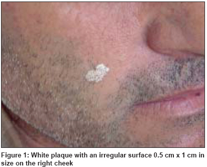
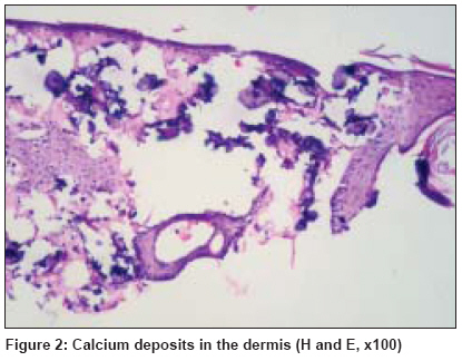
Indian Journal of Dermatology, Venereology and Leprology, Vol. 75, No. 2, March-April, 2009, pp. 180-181 Images in Clinical Practice Calcinosis cutis on the face Kayhan TubaCelebi, Temiz Peyker, Ermertcan AylinTurel Department of Dermatology, Celal Bayar University, Faculty of Medicine, Manisa Code Number: dv09050 A 46-year-old male patient came to our outpatient clinic with a white lesion on his right cheek. He explained that this lesion occured approximately 25 years ago. There was no family history of similar lesion. Dermatological examination revealed white plaque with an irregular surface 0.5 cm x 1 cm in size on his right cheek, which seemed to be whitewashed [Figure - 1]. Skin biopsy was performed from the lesion. Histopathological examination of the specimen revealed calcium deposits in the dermis [Figure - 2]. We diagnosed the patient as calcinosis cutis. Laboratory investigations revealed normal calcium and phosphate levels. Other laboratory tests for systemic diseases were also in normal ranges. Calcinosis cutis is a term used to describe a group of disorders in which calcium deposits form in the skin. In general, multiple, firm, whitish dermal papules, nodules or subcutaneous nodules are found. There are many classifications of the disease but commonly calcinosis cutis is divided into four groups: dystrophic, metastatic, iatrogenic and idiopathic. The dystrophic form is the most commonly described, whereas the idiopathic is the rarest. Dystrophic calcinosis includes those conditions in which calcification occurs in the damaged tissue. Various conditions can cause dystrophic calcinosis, including connective tissue disease, infection, inflammatory processes, chronic venous stasis, cutaneous neoplasm and trauma. This may be localized (calcinosis circumscripta) or widespread (calcinosis universalis). Metastatic calcification refers to deposition of calcium resulting from elevated serum levels of calcium or phosphorus. It is often associated with bone loss or destruction, the bone providing the source of the elevated serum calcium. Conditions associated with metastatic calcinosis include parathyroid neoplasms, primary hyperparathyroidism, hypervitaminosis D, sarcoidosis and extensive intake of milk and alkali. The most common metabolic condition associated with metastatic calcification is renal failure. Iatrogenic and traumatic calcinosis is associated with medical procedures or occupational exposures that may involve both tissue damage and local elevated calcium concentrations. Idiopathic calcinosis cutis refers to those forms of cutaneous calcification of unknown cause with normal serum calcium. Idiopathic scrotal calcinosis, subepidermal calcified nodule and tumoral calcinosis are idiopathic forms of calcification. Subepidermal calcified nodule is an uncommon but distinct type of idiopathic calcinosis. It occurs more commonly in children and can be present at birth. Clinical presentation generally consists of a single, white-yellowish small papule but multiple lesions can be seen less often. Most lesions occur on the face. According to some writers, calcinosis circumscripta are found in generalized scleroderma or dermatomyositis, but it may rarely occur as an idiopathic disorder. Our patient had no history of trauma and his lesion had occurred in childhood. His laboratory investigations revealed normal calcium and phosphate levels. Although our patient was mimicking idiopathic calcinosis cutis due to his history, clinical appearance of the lesion did not resemble any type of calcinosis cutis. Our case is very rare and interesting, with its unique clinical presentation. We suggest that calcinosis cutis should be kept in mind while thinking of non-specific and asymptomatic white plaque on the face, the classification of which may be looked over otherwise. Copyright 2009 - Indian Journal of Dermatology, Venereology and Leprology The following images related to this document are available:Photo images[dv09050f2.jpg] [dv09050f1.jpg] |
| |||||||||
